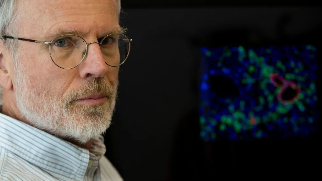
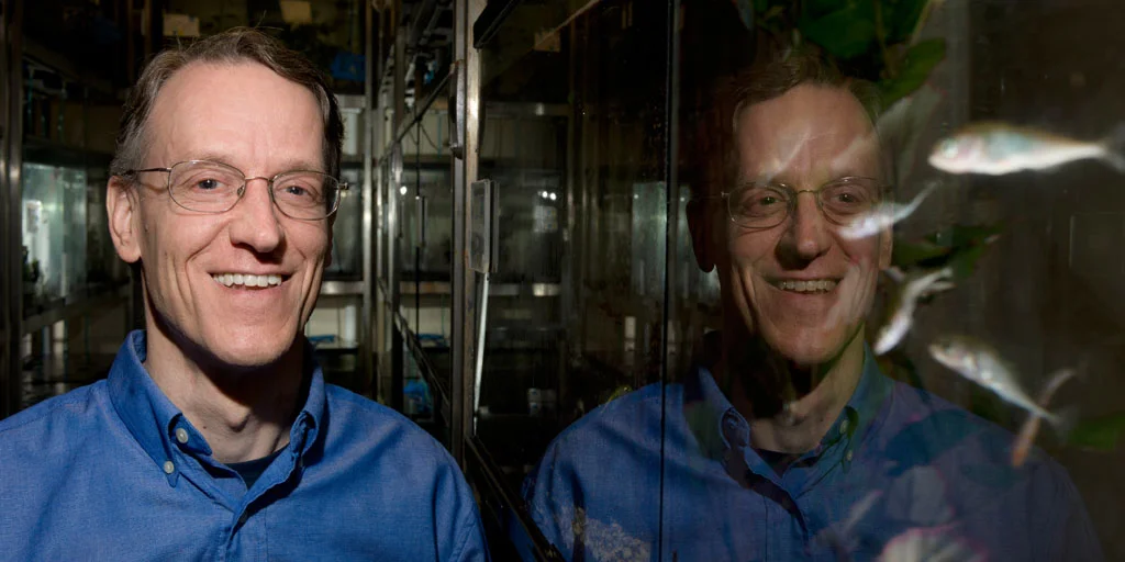
Defect in pancreas alpha cells linked to diabetes, Stanford Medicine study shows
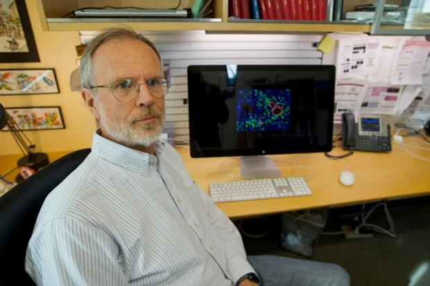
Pancreatic alpha cells from people with diabetes release excess amounts of glucagon, a hormone important in blood sugar control, in a new Stanford-developed mouse model of transplanted human islets.
In response to low blood sugar levels, pancreatic islet cells from people with diabetes release significantly more of a hormone called glucagon than do islet cells from healthy people, according to a new study by researchers at the Stanford University School of Medicine.
The discovery was made using a new mouse model of diabetes engineered by Stanford scientists that for the first time permits functional studies of transplanted human alpha cells.
Glucagon is produced by alpha cells in pancreatic islets while insulin is produced by beta cells. Defects of insulin output and beta cells have been thought to be the main drivers of diabetes. The current study, however, supports the growing realization that diabetes is likely due to defects in multiple cell types and highlights the importance of the mouse model to more accurately simulate the complexities of the disease.
“Our investigations support the idea that diabetes results not just from a defect in pancreatic beta cells, but also in the alpha cells that produce glucagon,” said Seung Kim, MD, PhD, professor of developmental biology and director of the Stanford Diabetes Research Center. “This had been suspected, but our study with human islets provides new, unprecedented evidence to support this idea.”
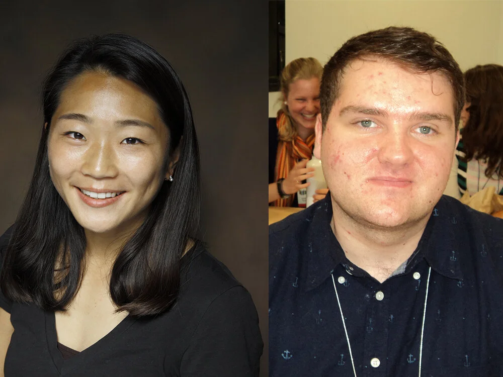
Kim is the senior author of the study, which was published June 8 in Nature Metabolism. Graduate student Krissie Téllez and research scientist Yan Hang, PhD, share lead authorship of the study.
In healthy people, glucagon works in tandem with insulin to tightly control blood sugar levels. Immediately after a meal, insulin triggers the removal of glucose from the blood and promotes its storage in organs like the liver. Conversely, when blood sugar levels become low, glucagon stimulates the release of stored glucose into the bloodstream.
Identical glucagon structures
Previous research into the relationship between insulin and glucagon has been hampered by the fact that mouse and human glucagon are identical in structure. They are impossible to distinguish from one another after human islets have been transplanted into laboratory mice. As a result, researchers could only study human glucagon production by alpha cells in test tubes or culture dishes, which does not accurately mimic what happens in the body.
Hang, Téllez and their colleagues set out to engineer a mouse strain unable to make its own glucagon. They used a genetic editing process called CRISPR to remove a small piece of the animal’s DNA that encodes for the glucagon protein. The process was laborious and time-consuming, taking more than two years to complete.
Eventually they produced mice with a combination of six specific mutations, including those necessary to allow the animals’ immune systems to accept transplantation of human pancreatic islets, and those enabling the study of human glucagon production in the animals.
“It was quite a testimony to the power of modern genetics and molecular biology,” Kim said. But the results were worth the effort, he added.
“When we compared islet cells from healthy and diabetic donors in test tubes, we saw some differences in glucagon secretion, but the results were not statistically compelling,” Kim said. “It really wasn’t clear what was going on. But once the human islets were engrafted into the mice, we could very clearly see a significant and sustained excessive output of glucagon from human islets when blood sugar levels drop. As a result, the blood glucose levels of these mice were higher than in mice transplanted with normal human islets.”
‘A game-changing moment’
Interestingly, the insulin production by the diabetic islets remained unchanged, indicating an additional defect in the beta cells’ response to the increased blood sugar levels. Kim and his colleagues are eager to find out why.
“The potential role of glucagon output and high blood sugar levels in diabetes has long been debated in diabetes research,” Kim said. “Now we can finally assess the interplay of islet cell types in people with and without diabetes and measure the role of multiple hormones involved in blood sugar control. These mice open up many new avenues of study, including the genetics and pharmacology of the disease and possible treatments. It really is a game-changing moment in diabetes research.”
Other Stanford co-authors are research associate Xueying Gu and postdoctoral scholar Charles Chang, PhD. A researcher from Vanderbilt University also contributed to the study.
The research was supported by the National Science Foundation, the National Institutes of Health (grants DK107507, DK108817, CA21192701, DK120447, DK106755, DK050203, DK090570 and DK116074), the HL Snyder Foundation, the Mulberry Foundation, S. and M. Kirsch, and the Stanford Diabetes Research Center.
By KRISTA CONGER
Krista Conger is a science writer in the Office of Communications. Email her at kristac@stanford.edu.
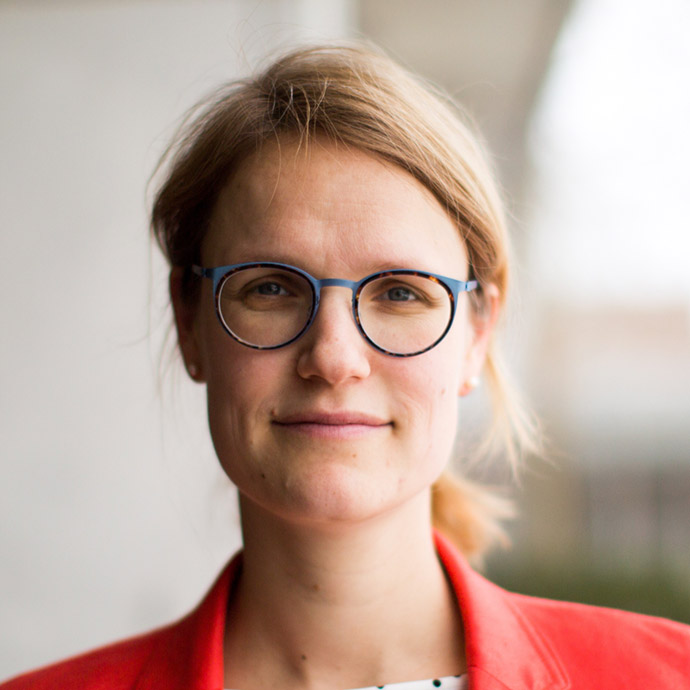
Emily Kolenbrander successfully defensed her here thesis, “Spatial and temporal constraints imposed on the Hedgehog pathway by the primary cilium”
On May 7, 2020, Emily Kolenbrander successfully defended her here thesis, “Spatial and temporal constraints imposed on the Hedgehog pathway by the primary cilium”
On May 7, 2020, Emily Kolenbrander successfully defended her here thesis, “Spatial and temporal constraints imposed on the Hedgehog pathway by the primary cilium”
Kelsey Roberts defended her thesis titled "Mechanism of Smoothened Regulation by the Hedgehog Receptor Patched"
On April 16, Kelsey Roberts defended her thesis titled "Mechanism of Smoothened Regulation by the Hedgehog Receptor Patched".
On April 16, Kelsey Roberts defended her thesis titled "Mechanism of Smoothened Regulation by the Hedgehog Receptor Patched”. Kelsey will soon start to work as a patent attorney.
Ellen Rim and Michael Guernsey successfully defended their thesis
During this academic year, several Developmental Biology graduate students successfully defended their thesis: Ellen Rim and Michael Guernsey.
During this academic year, several Developmental Biology graduate students successfully defended their thesis: Ellen Rim and Michael Guernsey. Ellen will soon start postdoctoral research in the area of plant biology. Michael will become a postdoc at UCSF, doing research on functional genetics and genomics to understand the evolution of bats.
One of our incoming students, Sherry Zheng has been awarded the NSF Graduate Research Fellowship.
Roel Nusse recognized with 2020 Gairdner Prize
Huge congratulations to Roel Nusse and members of his laboratory, who are are being recognized for the identification of Wnts and the elucidation of the Wnt pathway and its key role in development, stem cells, and cancer.
The 2020 winners of the prestigious Gairdner Award were announced today:
https://gairdner.org/winners/current-winners/
Huge congratulations to Roel Nusse and members of his laboratory, who are are being recognized for the identification of Wnts and the elucidation of the Wnt pathway and its key role in development, stem cells, and cancer.
Janet Rossant wrote a nice overview of the interrelated science themes behind the entire collection of 2020 awardees here:
https://www.cell.com/cell/pdf/S0092-8674(20)30289-0.pdf
It’s great to see this recognition for key developments in the basic science of cell and developmental biology.
I will also point out that Lucy Shapiro was honored with a Gairdner Award in 2009. Isn’t it wonderful to work in a department led by amazing scientists whose work is clearly shaping the entire field of developmental biology?
On behalf of all of us, congratulations and keep up the good work!
David Kingsley, Ph.D.
Professor and Associate Chair of Developmental Biology
HHMI and Stanford University School of Medicine
Stanford, CA. 94305-5329
Stanford’s response to COVID-19 (coronavirus)
Stanford continues to take steps to respond to the spread of coronavirus, working to support the health and well-being of our community and our neighboring communities.
Stanford continues to take steps to respond to the spread of coronavirus, working to support the health and well-being of our community and our neighboring communities. In-person instruction has been suspended, undergraduates who are able to do so have been asked to return to their homes, and the university is complying with new “shelter-in-place” orders issued for the Bay Area. Information on other impacts is available throughout this website. For daily email updates, please sign up using the “Email Alerts” box at https://healthalerts.stanford.edu/.
Stem cell researcher Irving Weissman awarded Albany Prize
Weissman and Johns Hopkins’ Bert Vogelstein will share the 2019 Albany Medical Center Prize in Medicine and Biomedical Research for discoveries in stem cell and cancer biology.
Irving Weissman, MD, director of Stanford’s Institute for Stem Cell Biology and Regenerative Medicine, will receive the 2019 Albany Medical Center Prize in Medicine and Biomedical Research for his pioneering work in stem cell and cancer biology, including the identification of blood-forming stem cells and their role in blood cancers, as well as the discovery of a “don’t eat me” signal on the surface of many cancer cells that protects them from being eliminated by the immune system.
Weissman is a professor of pathology and of developmental biology at the Stanford School of Medicine and is the director of the Ludwig Center for Cancer Stem Cell Research at Stanford. He will share the $500,000 prize with Bert Vogelstein, MD, who is the Clayton Professor of Oncology and Pathology at Johns Hopkins University’s Sidney Kimmel Cancer Center and the director of its Lustgarten Laboratory for Pancreatic Cancer Research.
Vogelstein is known for discovering that a protein called p53 functions as a tumor suppressor and that its inactivation is critical to the development of many human cancers. He was also the first to demonstrate in colorectal cancer that disease progression is a multistep process resulting from the sequential accumulation of mutations in specific cancer-associated genes. Together, Weissman and Vogelstein transformed the understanding of cancer biology, cancer genomics and disease initiation and progression, paving the way for earlier diagnosis and more effective treatments for a wide range of diseases including leukemia, non-Hodgkin lymphoma and severe combined immunodeficiency (also known as “bubble boy” disease).
The two will be presented with the prize at a Sept. 25 ceremony in Albany, New York.
“Dr. Weissman’s groundbreaking work in advancing our understanding of blood-forming stem cells and cancer has transformed many aspects of modern medicine,” said Lloyd Minor, MD, dean of the Stanford School of Medicine. “The discovery of the ‘don’t eat me’ signal on cancer cells promises to lead to novel clinical applications that will improve human health. We congratulate Dr. Weissman on this well-deserved recognition.”
Adult stem cells are unique in that they can both self-renew and make progenitor cells that give rise to all the specific cell types in a particular tissue of the body. In 1988, Weissman was the first to identify and isolate in mice the hematopoietic, or blood-forming, stem cells that form all the cells of the blood and immune system. In 1992, he and his group found the human blood-forming cells. He and his group have since painstakingly traced the cellular steps leading from a stem cell to each of the many types of mature blood and immune cells in humans, and identified those that go awry in many blood diseases and cancers.
Weissman also identified a molecule called CD47 that exists on the surface of nearly every human cancer cell and protects them from attack by immune cells called macrophages. An antibody targeting CD47, which the researchers have termed a “don’t eat me” signal, is in clinical trials in people with several types of blood and solid cancers. Overexpression of CD47 is also implicated in fibrotic diseases such as scleroderma and surgical adhesions. Recently, Weissman identified additional “don’t eat me” signals, each of which is expressed by particular types of cancers.
“I’m especially honored to share this award with Bert Vogelstein, whose work I have followed for many years and greatly admire,” said Weissman, who is the Virginia and D.K. Ludwig Professor for Clinical Investigation in Cancer Research. “Inspired by his earlier work on colon cancer, we were able to show that nearly all stepwise mutations that lead to the development of leukemia and blood diseases, such as myelodysplastic syndrome, occur in blood-forming stem cells, apparently hitchhiking in these self-renewing cells to form disease clones. It’s a fantastic feeling to join the group of highly accomplished past recipients of the Albany Prize.”
The Albany Prize is funded by a $50 million gift from New York City philanthropist Morris Silverman. It has been awarded since 2001 to encourage and recognize extraordinary and sustained contributions to improving health care and promoting biomedical research with translational benefits for better patient care. In 2015, Karl Deisseroth, MD, PhD, Stanford professor of bioengineering and of psychiatry and behavioral sciences, received the award.
Genetics Society of America Awards 2019 GSA Medal to Anne Villeneuve
The Genetics Society of America (GSA) is pleased to announce that Anne Villeneuve, PhD, of Stanford University is the recipient of the 2019 Genetics Society of America Medal.
The Genetics Society of America (GSA) is pleased to announce that Anne Villeneuve, PhD, of Stanford University is the recipient of the 2019 Genetics Society of America Medal. Villeneuve is recognized for her research on the mechanisms governing chromosome inheritance during sexual reproduction. Her research focuses on meiosis, the specialized cell division program involved in generating egg and sperm cells. Meiosis enables diploid organisms (which have two copies of each chromosome) to generate haploid gametes (which have only a single set of chromosomes). This halving of chromosome number is crucial for sexual reproduction, as it allows restoration of the diploid chromosome number in the offspring formed once the egg and sperm fuse.
As an independent fellow at Stanford, Villeneuve recognized the considerable untapped potential of the nematode Caenorhabditis elegans as an experimental system for studying chromosomes during meiosis. Trained as a geneticist, she set out to exploit this opportunity by conducting screens to identify genes important for meiosis, most famously an elegant approach nicknamed “Green eggs and Him,” which was published in GENETICS and continues to be used as an exemplar in many university genetics courses.
Research from Villeneuve’s lab and those of her former trainees has played a key role in establishing C. elegans as one of the premier experimental systems for investigating chromosome organization, genetic recombination, and genome maintenance in the context of meiosis. Villeneuve’s research integrates sophisticated genetic strategies with high-resolution and super-resolution cytological imaging of chromosomes in the context of an optically transparent gonad in which germ cells progressing through meiosis are arranged in a temporal/spatial “time course.” This approach has enabled them to identify numerous components of the machinery responsible for the key chromosomal and DNA events of meiosis and to elucidate the mechanisms underlying these events and how they are coordinated. A substantial fraction of the genetic mutants, assays, and cytological reagents used to investigate genome maintenance, recombination, and meiosis in C. elegans was developed in her laboratory.
Papers published by the Villeneuve lab during the past 15 years have had a significant impact on our understanding of most major aspects of the meiotic program, including pairing between homologous chromosomes; structure, function, assembly and dynamics of the synaptonemal complex (SC), a meiosis-specific structure located at the interface between aligned homologs; formation and repair of DNA double-strand breaks; spatial patterning and maturation of meiotic crossovers; remodeling of chromosome structure in response to recombination; regulated release of sister chromatid cohesion; organization of chromosomes on the oocyte meiotic spindle; and quality control mechanisms that ensure a robust outcome of meiosis.
“Literally every major event in meiosis has been dissected in Anne’s lab and generated beautiful, high-profile papers,” says Barbara Meyer, a Professor at the University of California, Berkeley and one of the scientists who nominated Villeneuve for the Medal. “It is extremely rare to be able to cite a single lab with such a huge impact on a field of biology.”
A hallmark of research from the Villeneuve lab is the generation of microscopic images of meiotic chromosomes that provide a stunning visual readout of the inner workings of meiosis. This is beautifully illustrated in a recent paper from postdoctoral researcher Alex Woglar, which revealed the distinct spatial architecture of recombination proteins localized at meiotic crossover sites and showed that recombination site architecture undergoes dynamic changes during meiotic progression.
Villeneuve’s group has also provided insight into the process of crossover interference, which was originally described by Muller over 100 years ago, yet has remained largely mysterious during the intervening century. Crossover interference refers to the non-random placement of crossovers, such that a (nascent) crossover reduces the likelihood that another crossover will be formed nearby. A series of papers from the Villeneuve lab exploited genetic and cell biological tools available in the worm to implicate the SC as an important conduit of communication along the chromosomes. Their findings support a model of meiotic crossover regulation as a self-limiting system in which SC components initially promote the formation of crossover intermediates, which in turn trigger a change in the state of the SC that inhibits further crossover formation.
“Anne is a true scholar and has made a number of significant and impactful contributions to the field of genetics over the last 15 years,” says JoAnne Engebrecht, a Professor at the University of California, Davis and one of the scientists who nominated Villeneuve for the GSA Medal. “She embodies the ingenuity of the GSA membership in using genetics to investigate a fundamental biological process.”
Villeneuve’s influence on the fields of meiosis and recombination in particular, and genetics in general, extends well beyond the research conducted in her own lab. She has an outstanding record for mentoring younger scientists, and many of her trainees have gone on to establish their own productive independent research groups. Moreover, she has fostered a collegial community among meiosis researchers and has provided mentorship for numerous scientists outside her own group. Villeneuve also has a long-standing involvement with the GSA. She first joined the GSA as a graduate student, served as its Secretary from 2013 to 2015, and was an Associate Editor for the GSA journal GENETICS from 2004 to 2010. Villeneuve’s scientific contributions and leadership in the Meiosis and Recombination fields have been recognized in recent years by a Research Professor Award from the American Cancer Society and by her election to the American Academy of Arts and Sciences in 2016 and the National Academy of Sciences of the USA in 2017.
The GSA Medal was established in 1981 to recognize members who have made outstanding contributions to the field of genetics during the past 15 years. The award will be presented to Villeneuve at the 22nd International C. elegans Conference, which will be held June 20–24, 2019 in Los Angeles, CA.
In the Spotlight: Working toward pure populations of stem cells
Stem cells give rise to a world of potential. Embryonic stem cells, for instance, can specialize into nearly any cell in the body: from blood, to brain, to muscle, opening doors for therapeutic applications. The problem, however, is that it's tricky to coax these cells to mature into a specific cell type.
Stem cells give rise to a world of potential. Embryonic stem cells, for instance, can specialize into nearly any cell in the body: from blood, to brain, to muscle, opening doors for therapeutic applications. The problem, however, is that it's tricky to coax these cells to mature into a specific cell type.
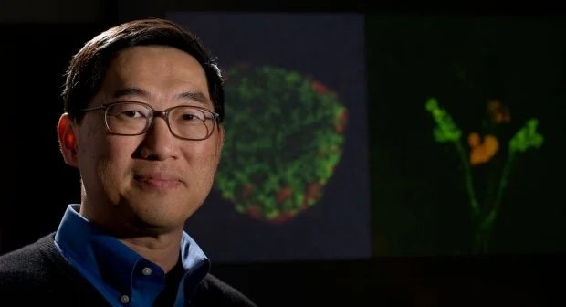
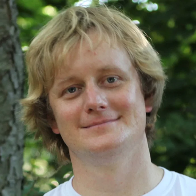
That's where researcher Kyle Loh, PhD, comes in. Originally from New Jersey, he came to Stanford in 2011, earned a PhD, and is now an assistant professor of developmental biology.
I spoke with him to learn more about his research and life in science.
What is your research focus?
My lab's work now extends from my PhD focus on embryonic stem cells. The work I'm doing now has been how to figure out how to turn embryonic stem cells into pure populations of different types of cells.
What have been some outcomes of your research?
We have made tremendous progress. For instance, we've been able to make nearly pure populations of human liver cells in a petri dish, and then inject these cells into a mouse, and we've seen the regeneration of liver tissue that partially rescues a mouse from liver failure. We've also made human bone cells that, when injected into a mouse, regenerate bone and cartilage.
Why did you go into science?
When I was in kindergarten, I read a science encyclopedia. In first grade, I read a book about the human body, through which I became fascinated by the immune system. Then, second grade, I read a book on Ebola virus, and that's really how I got into things in the realm of science.
I was also very lucky that at around age 13, I had the unusual opportunity to take courses in community college. I graduated at age 16 with my bachelor's from Rutgers University, the state university of New Jersey.
What's better -- East Coast or West Coast?
There's definitely a lot more sunshine here. But a part of the Bay Area and Stanford that has stood out to me is the collaborative atmosphere. A lot of the work that I've talked about was uniquely enabled because I could collaborate with other Stanford scientists, who are experts in those areas. It's been a huge amount of fun.
What does a normal day look like for you?
It definitely varies. Since I recently started my faculty career, I spend a lot of time in the lab working on experiments. I also dedicate a lot of time to reading scientific literature, teaching, writing grants, as well as mentoring students in my lab. I really value the dynamic of my lab -- it's comprised of only six of us and it's a very tight knit group.
What are you looking forward to?
I'm really excited about a current experiment in which we are hoping to generate new human blood in a petri dish from embryonic stem cells. This is really exciting because we can make all kinds of blood cells patients need, like red blood cells, platelets, and special immune cells called T cells. Being able to mass produce T cells, for instance, would be very useful in treating cancer and for immunotherapy.
How do you unwind?
I enjoy road biking, which is a hobby I picked up at Stanford. A number of colleagues and I go road biking on the weekends, which is a lot of fun, and the scenery around Stanford is perfect for it.
What are you reading now?
I most recently read Ahead of the Curve, a book about the Nobel Laureate David Baltimore, PhD, and his life in science. It shows he was a very, very intense person, especially in his younger days when he made most of his discoveries.
What are your favorite foods?
That's an easy one: steak. Specifically prime rib.
Do you have a mantra you live by?
Whatever you want to do in life requires your full commitment, so that whatever you set your mind to, you can do it well.
Photo by Chris Vaughan
Four faculty members appointed to endowed professorships
Timothy Cornell, Kevin Shea, Joanna Wysocka and Tony Wyss-Coray have been appointed to endowed professorships.
Timothy Cornell, MD, professor of pediatrics, was appointed the Chambers-Okamura Endowed Professor of Pediatric Critical Care Medicine, effective Dec. 4. His research interests include the role of epigenetics in the regulation of inflammation and the use of molecular biomarkers and precision medicine to improve outcomes for critically ill children.
The professorship was established with a gift from Jeffrey Chambers and Andrea Okamura to support the division chief of critical care in the Department of Pediatrics. Chambers, who earned an MBA from Stanford, is chair of the board of directors at Lucile Packard Children’s Hospital Stanford.
Kevin Shea, MD, professor of orthopaedic surgery, was appointed the Chambers-Okamura Endowed Professor of Pediatric Orthopaedics, effective Dec. 4. His research interests include health care system performance and quality improvement, sports medicine, ACL injury and the treatment of osteochondritis dissecans of the knee, elbow and ankle.
The professorship was established with a gift from Jeffrey Chambers and Andrea Okamura to support a faculty member with a focus on pediatric orthopaedics in the Department of Orthopaedic Surgery.
Joanna Wysocka, PhD, professor of chemical and systems biology and of developmental biology, was appointed the Lorry Lokey Professor, effective Dec. 4. Her research examines how gene regulation and expression are related to human development, evolution and disease.
The professorship was established with a gift from Lorry Lokey and an anonymous donor to support a basic science faculty member who specializes in stem cell research. Lokey, a Stanford alumnus and significant donor, founded Business Wire, which distributes press releases and regulatory disclosures.
Tony Wyss-Coray, PhD, professor of neurology and neurological sciences, was appointed the D.H. Chen Professor II, effective Dec. 4. His research focuses on brain aging, neurodegeneration and Alzheimer’s disease.
The professorship was established with funds from the D.H. Chen Foundation and an anonymous donor to support a School of Medicine faculty member in the neurosciences.
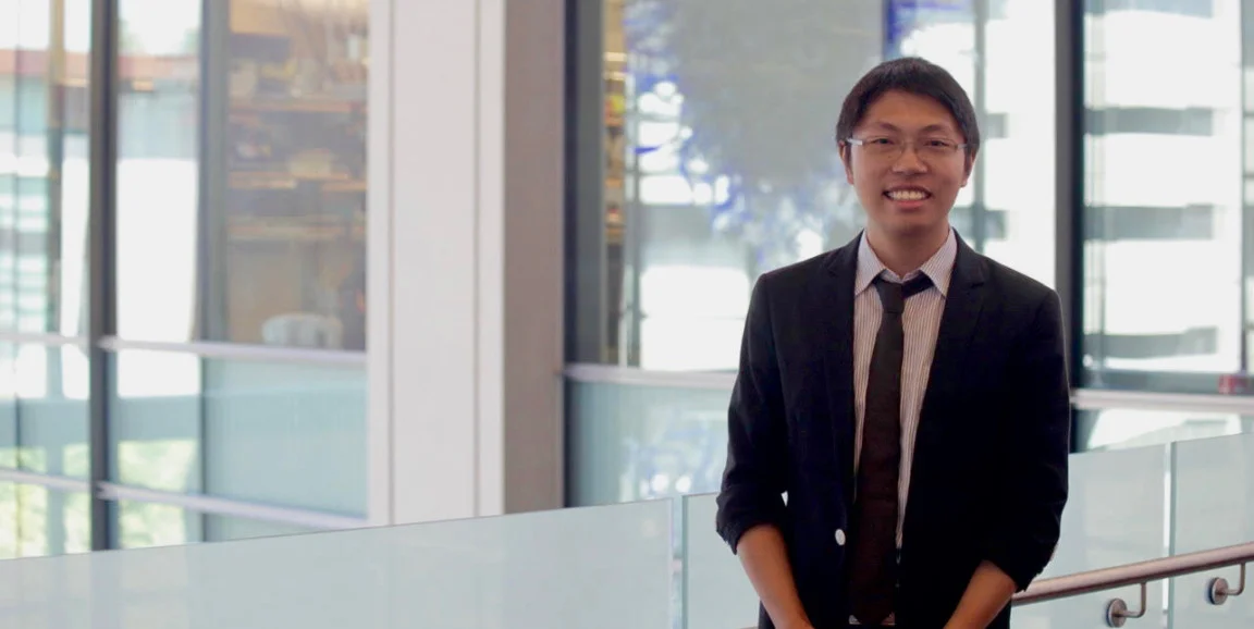
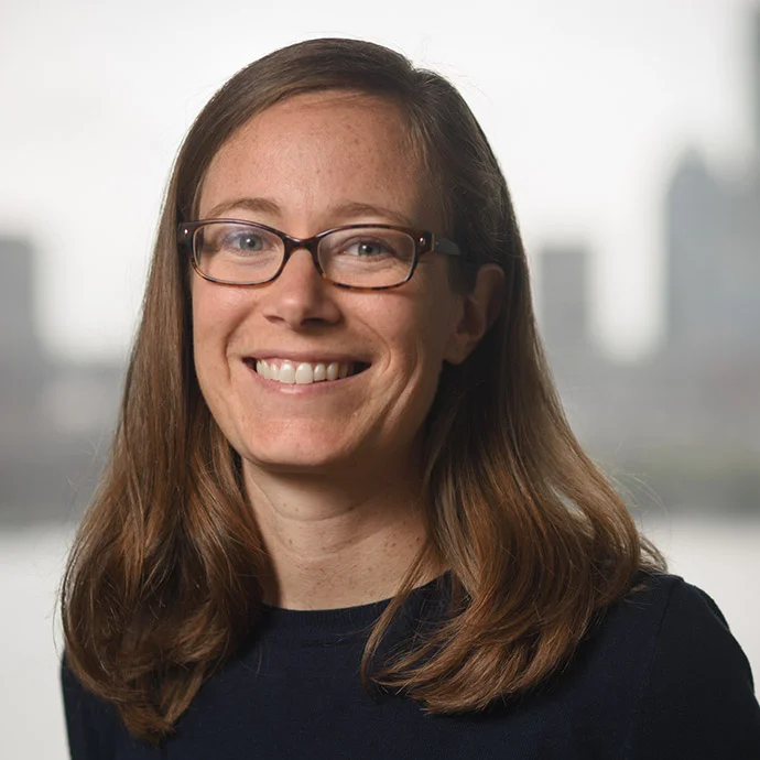
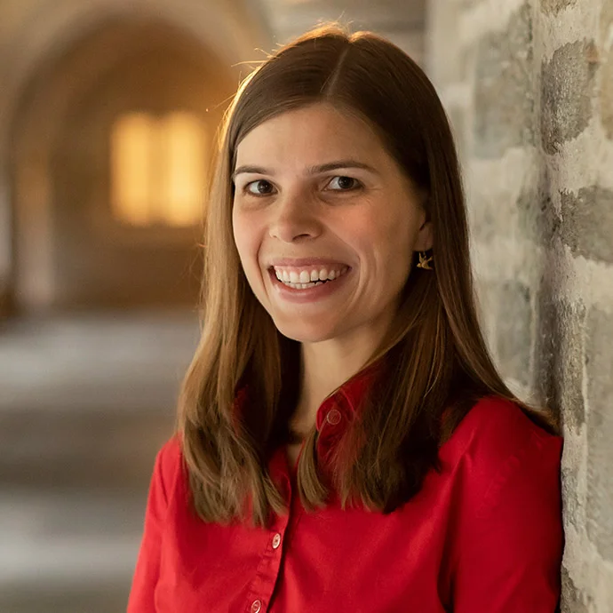
In the Spotlight: Hormone regulation and science fiction
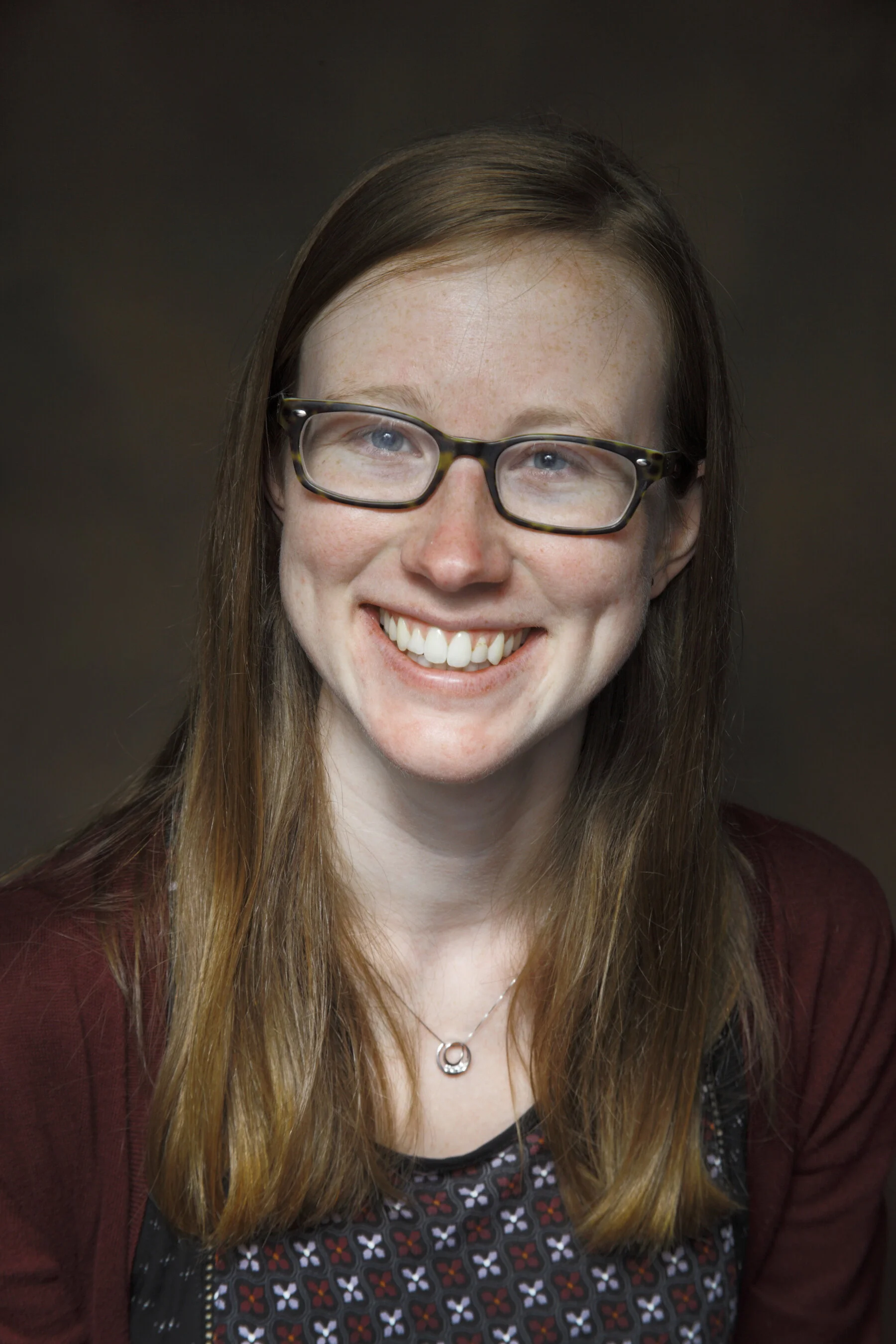
When she arrived at San Francisco State to study cell molecular biology as an undergrad, Krissie Tellez was convinced the Bay Area topped her native Southern California.
When she arrived at San Francisco State to study cell molecular biology as an undergrad, Krissie Tellez was convinced the Bay Area topped her native Southern California. Now, in her fifth year as a developmental biology graduate student, she's leaning a bit more toward Los Angeles. "I miss being able to go to the beach whenever I want," Tellez admitted.
When I caught up with her recently, we geeked out about science fiction and her research — here's a closer look:
Why did you go into science?
I have no scientists in my family. It was something I ventured into on my own.
In high school, and still to this day, I was a total sci-fi nerd, so when I was in my high school biology class and my teacher mentioned stem cells and their potential to go from blank cells and into mature cells, I was super fascinated.
What are you working on today?
My current project focuses on regulation of a hormone called glucagon. It’s secreted from alpha cells in your pancreatic islets of Langerhans, and acts opposite of insulin. Glucagon tells your liver to release any glucose stores and generate new glucose while you're fasting, like during sleep. It also plays a role in diabetes. We see that in patients with diabetes not only does insulin decrease, but glucagon is misregulated.
What is your daily routine like?
I’ll come in, check my emails, and see if there are any human tissue donations for our experiments. I check on my mice.
Then, it depends. Some days I’m really busy running experiments like Elisas, which are plates coated with antibodies where we put mouse serum or media from cell culture and measure small amounts of proteins, like glucagon. Other days I spend a lot of time with my mice. About once a week, I like to have a more relaxed day where I'm able to spend a majority of my time reading and/or writing.
What is a challenge in your field right now?
For my field of research, the number of islets (a part of the pancreas) is a challenge, so it’s a quantity problem. From one mouse, you can maybe get 150-400 islets at most, so analyzing certain interactions is very tough. Also, it would be great if we could keep islets alive in culture for longer. Right now, I’m usually limited to about a week, because after that the natural regulated secretions of the hormones usually become disrupted.
What is most fulfilling about your work?
There’s this really fun discovery aspect that I feel is very specific to science. Having a question and being able to test that question experimentally and work toward answers — it’s fun.
What sci-fi films or books can you recommend?
I really love the 'Star Wars' franchise. As far as novels, 2001: A Space Odyssey is so beautifully written and one of my all time favorites.
What do your colleagues not know about you?
I make clothes. I love sewing, it’s been a nice way to relax.
Any advice for future scientists?
The biggest advice is to take ownership of your research projects. Start research as early as possible. Find a PI that’s interested and supportive of you — those relationships are really important.
What do you want to learn or know how to do that you don’t now?
I’d like to learn coding — especially R. I wish I had learned in undergrad, or earlier in grad school, because data analysis of sequencing data does requires some knowledge of these coding languages.
Do you have a mantra you live by?
Be truthful, especially in science. It’s okay for things to fail.
What is your ultimate career goal?
I’m still considering a lot of things, including science policy. I like the idea of being able to advocate for scientists.
I’m also looking into science communication. I think it’s important to take what we know as scientists and translate it for the public.
Academia is also an option. I was lucky to have amazing mentors, so I’d love to be a part of inspiring young scientists and support them.
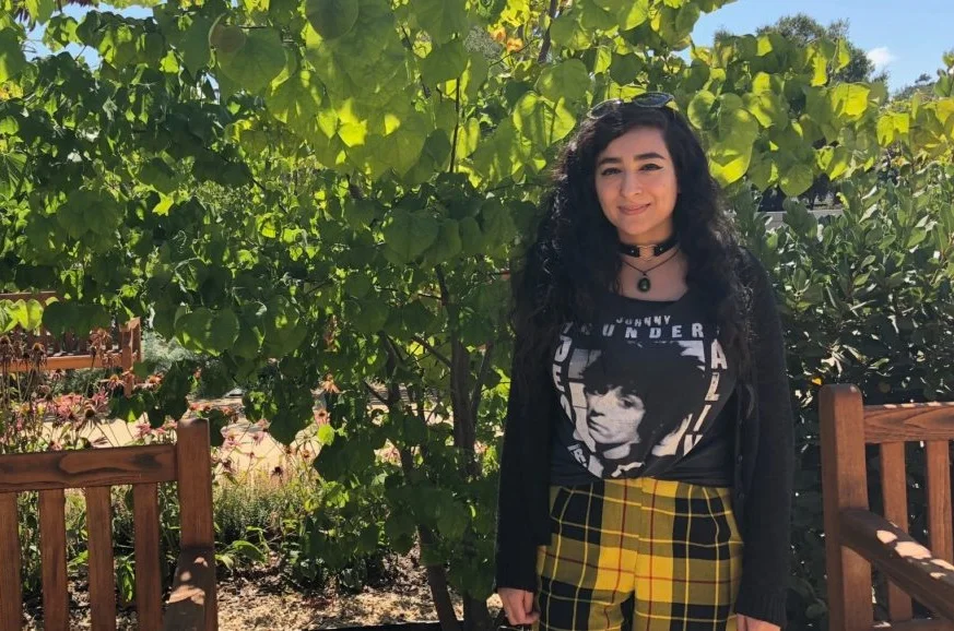
By Roxanne Ohayon
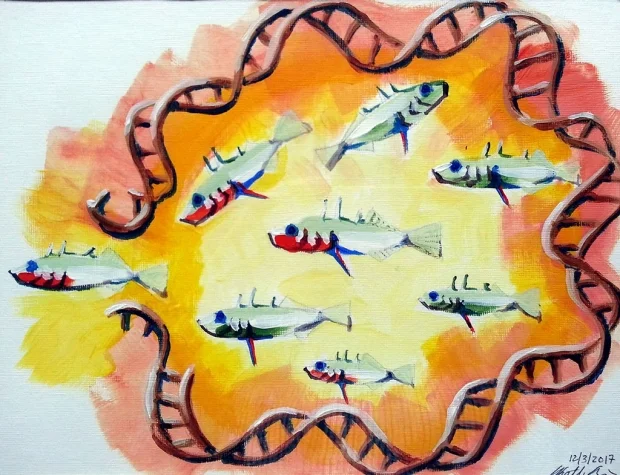
Strength in weakness: Fragile DNA regions key to vertebrate evolution
DNA regions susceptible to breakage and loss are genetic hot spots for important evolutionary changes, according to Stanford study. The findings may lead to new understanding of human evolution.
Regions of DNA susceptible to deletion during replication may have allowed vertebrates to successfully adapt to rapidly changing environmental conditions during evolution, according to a study by researchers at the Stanford University School of Medicine.
The research suggests that some critical evolutionary changes are likely to have occurred in leaps and bounds through the abrupt loss of stretches of DNA, rather than through the slow accumulation and additive effects of many small mutations.
The researchers, who studied a tiny fish called the threespine stickleback, found that such “fragile” DNA regions create genetic hot spots that mutate much more rapidly, and dramatically, than neighboring sequences. The resulting changes can help an organism vault far ahead of its peers in the evolutionary arms race.
Although similar findings have been described in bacteria, this is one of the first studies to show that the same process has occurred in vertebrates to create dramatic changes in body structure. It also addresses a long-standing mystery in evolutionary biology.
“There is a lot of evidence that the same genes across different populations or species are often responsible for similar evolutionary changes,” said David Kingsley, PhD, professor of developmental biology. “What hasn’t been clear is why this is happening. This study describes at a biochemical level, down to the atoms and sequences in DNA, how a particular type of mutation can arise repeatedly, which then contributes to a complex skeletal trait evolving over and over again in wild fish species. It’s a great example of how DNA fragility can sometimes contribute to favorable traits rather than diseases in natural populations, and it may give us important insights into the process of human evolution.”
Kingsley, a Howard Hughes Medical Institute investigator, is the senior author of the study, which was published Jan. 4 in Science. Graduate student Kathleen Xie is the lead author of the work.
Large changes, large effects
Many mutations involve a change in just a single nucleotide, or letter, of DNA. Few of these “point” mutations will confer an evolutionary advantage on their own. Instead, significant change often requires the gradual accumulation of several such mutations. In contrast, sudden, large changes in the genome can have large effects — changing body structure through skeletal modifications or affecting metabolism or brain function, for example. Often, these changes are deleterious, decreasing the chances of an animal’s survival. Occasionally, however, the changes are advantageous.
David Kingsley
When the last Ice Age ended, about 10,000 years ago, pockets of migratory ocean threespine sticklebacks colonized newly formed lakes and streams in coastal regions, and then evolved independently in response to their new local environments. As a result, many of these populations show significant differences in body structure. Marine sticklebacks, for example, have a hind fin with a large spine projecting down from their pelvic structure. In contrast, dozens of freshwater populations have lost that hind fin; its absence likely reduces their need for calcium and chances of being nabbed and eaten by hungry insects.
Previous studies in the Kingsley laboratory have identified the loss of a specific DNA regulatory region, called the Pel enhancer, as the repeated cause of the missing hind fins in many populations of the freshwater fish. The Pel enhancer drives the expression of a protein necessary to trigger hind fin development. In this study, Xie used marine stickleback DNA to investigate the Pel region that is missing in its freshwater brethren to learn why that region was particularly susceptible to loss.
Xie found that the DNA sequence of the Pel region is unusual in several ways. Unlike surrounding regions, which exhibit the normal, more-stable helical twist associated with most DNA, the Pel enhancer region that was lost formed an alternate DNA structure predicted to be highly flexible and likely to be unstable during DNA replication. The sequence also contains long strings of repeated pairs of nucleotides, like a kind of genetic stutter. Previous studies in bacteria, mice and humans have indicated that these repeats are often associated with deletions of stretches of DNA.
More frequent chromosome breaking
When Xie tested the stability of the missing Pel region by inserting it into artificial yeast chromosomes, she found that the chromosome broke about 25 to 50 times more frequently than typical DNA sequences. When Xie and her collaborators then tested similar DNA sequences in mammalian cells, they observed that the key dinucleotide repeat sequence often led to the deletion of sections of DNA more than 100 nucleotides long.
The increase in the rate of chromosome breakage observed by Xie, coupled with the likelihood that this damage causes deletions of entire sections of DNA, may have been a key factor in allowing the prominent hind fin skeletal trait to emerge over and over again in many different young stickleback populations. Elevated mutation rates may play a similar role when advantageous traits arise in other organisms, the scientists believe.
“Many vertebrates, including early humans, are dealing with a small population size and relatively long generation times,” said Kingsley, who is the Rudy J. and Daphne Donohue Munzer Professor in the School of Medicine. “There aren’t that many generations available in which to evolve new, potentially advantageous traits. Under these conditions, it may be particularly important for mutations to occur at elevated rates, and to have sweeping effects.”
When the researchers investigated known instances of adaptive changes in humans, they found that about half were due to mutations that also arise at elevated rates compared with more typical DNA letter changes.
“What we’re learning is that ‘arrival of the fittest,’ or the relative speed with which a potentially favorable mutation arises, can sometimes be as important as ‘survival of the fittest,’” Kingsley said. “The mutation process itself has an important effect on the outcome, and the arrival of the mutation interacts with its effect on the fitness of the organism to bring about major changes in vertebrate evolution.”
Kingsley is a member of Stanford Bio-X and the Wu Tsai Neurosciences Institute.
Other Stanford authors are former graduate student Abbey Thompson, PhD, and graduate student Julia Wucherpfennig. Researchers from the University of Texas-Austin, the University of Victoria, the University of Nottingham, the University of British Columbia and the University of California-Berkeley also contributed to the study.
The research was supported by the National Institutes of Health (grants 5P50HG2568, CA093729 and 2T32GM007790), the National Science Foundation, a Stanford CEHG Graduate Fellowship and the Howard Hughes Medical Institute.
Stanford’s Department of Developmental Biology also supported the work.
By KRISTA CONGER
Krista Conger is a science writer for the medical school's Office of Communication & Public Affairs. Email her at kristac@stanford.edu.
Four faculty members appointed to endowed professorships
Andra Blomkalns, Gerald Grant, David Kingsley and Crystal Mackall have been appointed to endowed professorships.
Andra Blomkalns, Gerald Grant, David Kingsley and Crystal Mackall have been appointed to endowed professorships.
Andra Blomkalns, MD, professor and chair of emergency medicine, was appointed the Stanford Medicine Professor in Emergency Medicine, effective Oct. 8. Her academic work has focused on clinical innovation, and evaluating and improving the process for technology development and commercialization within medicine. She also has studied cardiovascular emergencies, obesity and dietary influences on health and disease.
The professorship was established in June using funds from the Department of Emergency Medicine, the School of Medicine’s Dean’s Office and Stanford Health Care.
Gerald Grant, MD, professor of neurosurgery, was appointed the Endowed Professor in Pediatric Neurosurgery, effective Oct. 16. He specializes in brain tumor and epilepsy surgery in children, and his laboratory is working to improve the delivery of drugs past the blood-brain barrier to reach brain tumors in children.
The professorship was established in 2015 to support a faculty member in pediatric neurosurgery. Funders include Jeffrey Chambers and Andrea Okamura; Roelof Botha and Huifen Chan; and the Schow Foundation.
David Kingsley, PhD, professor of developmental biology, was appointed the Rudy J. and Daphne Donohue Munzer Professor in the School of Medicine, effective Oct. 16. His research examines the molecular mechanisms that underlie evolutionary traits and common diseases in vertebrates.
The professorship was established in 1990. Rudy Munzer was the president and chairman of Petrolane, and Daphne Munzer volunteered with numerous Southern California organizations, including the Children’s Dental Health Clinic and the Long Beach Public Library.
Crystal Mackall, MD, professor of pediatrics and of medicine, was appointed the Ernest and Amelia Gallo Family Professor, effective Oct. 16. She is the founding director of the Stanford Center for Cancer Cell Therapy and directs the Parker Institute for Cancer Immunotherapy at Stanford. Her research focuses on enhancing the effectiveness of T cell-based cancer immunotherapies.
The professorship was established in October with a gift from the Ernest Gallo Foundation in honor of Ernest and Amelia Gallo, as well as matching funds from an anonymous donor. Ernest Gallo co-founded E&J Gallo Winery in 1933.
Stanford Medicine integrates research, medical education and health care at its three institutions - Stanford University School of Medicine, Stanford Health Care (formerly Stanford Hospital & Clinics), and Lucile Packard Children's Hospital Stanford. For more information, please visit the Office of Communication & Public Affairs site at http://mednews.stanford.edu.

Packard Fellowships in Science and Engineering Announces New Class of Fellows and Celebrates 30th Anniversary
Today, the David and Lucile Packard Foundation celebrated the 30th anniversary of the Packard Fellowships for Science and Engineering by announcing the recipients of its 2018 class of Fellows.
October 15, 2018 • In Science, Packard Fellowships for Science and Engineering
October 15, 2018 (Los Altos, CA) – Today, the David and Lucile Packard Foundation celebrated the 30th anniversary of the Packard Fellowships for Science and Engineering by announcing the recipients of its 2018 class of Fellows. Each of the 18 innovative, early-career scientists and engineers will receive $875,000 over five years to pursue their research.
The Packard Fellowships in Science and Engineering are among the nation’s largest nongovernmental fellowships, designed to allow maximum flexibility in how the funding is used. Since 1988, this program has supported the blue-sky thinking of scientists and engineers in the hopes that their research over time will lead to new discoveries that improve people’s lives and enhance our understanding of the universe.
The Packard Fellows’ work has contributed to breakthroughs like the creation of the CRISPR-Cas9 gene-editing technique, the discovery of soft tissues in dinosaur fossils, and the first-ever observation of a neutron star collision. Fellows have gone on to receive a range of accolades, including Nobel Prizes in Chemistry and Physics, the Fields Medal, the Alan T. Waterman Award, MacArthur Fellowships, and elections to the National Academies. The Fellows also gather at annual meetings to discuss their research, where conversations have led to unexpected collaborations across disciplines.
“It really is amazing to see what brilliant researchers can do when given the room to take big risks,” said Frances Arnold, Chair of the Packard Fellowships Advisory Panel, 2018 Nobel Laureate in Chemistry, and former Packard Fellow. “And I’m not only talking about their impressive contributions to their fields—I’m also talking about building entirely new disciplines and giving back to the next generation of scientists. I’m excited to see what’s in store for this new class as it joins our welcoming community of Fellows.”
The Fellowships program was inspired by David Packard’s commitment to strengthen university-based science and engineering programs in the United States. He recognized that the success of the Hewlett-Packard Company, which he cofounded, was derived in large measure from research and development in university laboratories. Since 1988, the Foundation has awarded $410 million to support 595 scientists and engineers from 54 national universities.
“At the Packard Foundation, we believe that every sector of society—from philanthropy to academia to government—has a crucial role to play in supporting science and research,” said David Orr, Chair of the Packard Foundation Board of Trustees. “Over the past three decades, the Fellowship program has been an example of our deep commitment to basic research in science and engineering. Every year, I love learning from the Fellows about their sometimes unexpected, but always fascinating, research—much of which would be difficult or impossible to perform with funds from traditional grants. The new class will certainly continue the tradition of the groundbreaking science that Packard Fellows have become known for.”
To commemorate 30 years of the Packard Fellowships, the Packard Foundation launched a new website exploring the inspiring work, ideas, and careers of 30 years of Fellows. Over 250 Fellows and their families also gathered this year to celebrate the program’s anniversary in southern California, where many Fellows had the chance to reflect on what the program has meant to them.
William Anderegg
Department of Biology, University of Utah
Discipline: Ecology, Evolutionary Biology
The future of Earth’s forests hangs in the balance between the potential benefits of rising atmospheric carbon dioxide and the stresses from climate change. Anderegg’s lab focuses on understanding and predicting the future of Earth’s forests using a mix of experiments, field measurements, and mechanistic models.
Michael Baym
Department of Biomedical Informatics, Harvard University
Discipline: Biological Sciences
Every time we have introduced an antibiotic, bacteria have rapidly evolved resistance. Baym’s lab uses a combination of experimental evolution and data science to understand this dynamic process, in order to design practical interventions to decrease, prevent, or even reverse resistance evolution.
Kristin Bergmann
Department of Earth, Atmospheric and Planetary Sciences, Massachusetts Institute of Technology
Discipline: Geosciences
From the landscape to the atomic scale, Bergmann and her group are reconstructing Earth’s ancient environmental conditions to explore the co-evolution of life and the Earth system. Her focus is on understanding climate dynamics during the radiation of complex life, in order to understand why it emerged on Earth—and to grasp how rare it may be in the Universe.
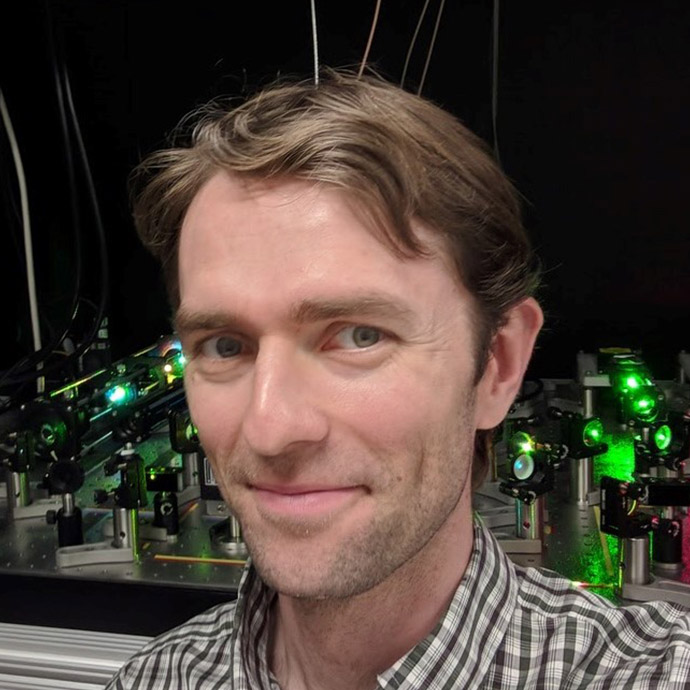
Alistair Boettiger
Developmental Biology, Stanford University
Discipline: Biological Sciences
Boettiger’s lab investigates the mechanism by which the three-dimensional structure of the genome is organized, and the consequences of this organization in regulating gene expression and in shaping cell fate specification during embryonic development. They pursue this with multiplex single-molecule imaging and transgenic techniques. Learn more about Boettiger’s work here.
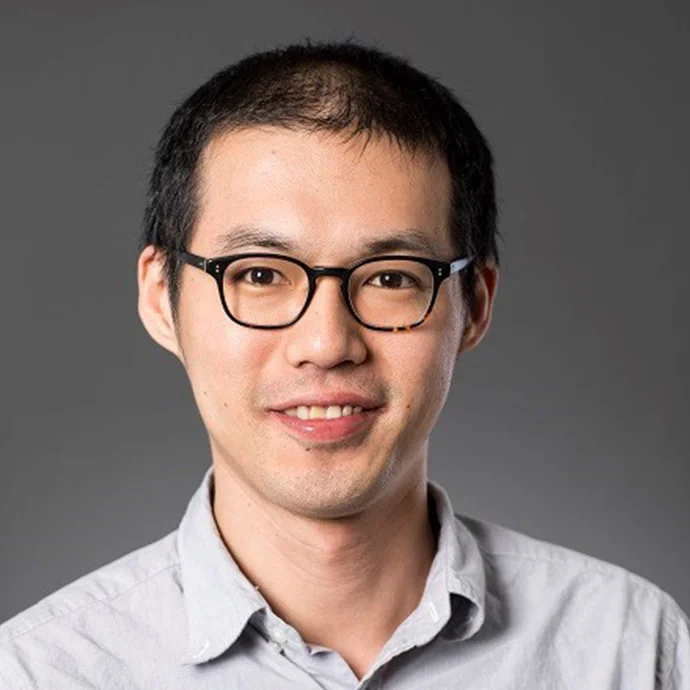
Jiun-Haw Chu
Department of Physics, University of Washington
Discipline: Physics
Chu’s lab aims to understand and eventually control the collective behaviors of quantum matter. They use material synthesis to create new model systems that help them attack open problems on multiple fronts, and they also design new experimental techniques to probe these materials in novel ways.
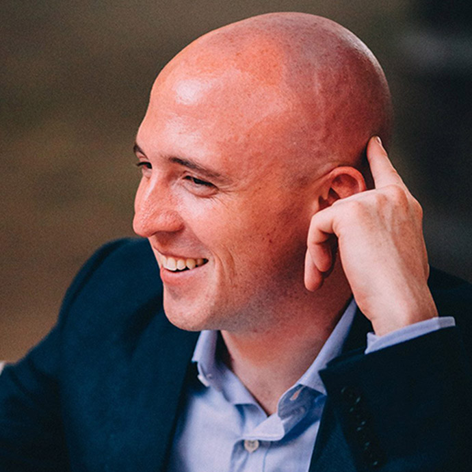
Keenan Crane
Computer Science Department, Carnegie Mellon University
Discipline: Computer/Information Sciences
Crane explores how the shapes and motions we observe in nature can be faithfully expressed in a language that is completely finite and discrete, and can hence be understood by a computer. His exploration of this question provides both new foundations for computation, as well as new ways to turn digital designs into physical, shape-shifting matter.
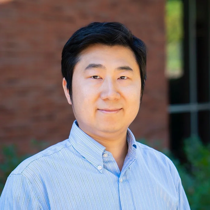
Weizhe Hong
Department of Biological Chemistry, University of California, Los Angeles
Discipline: Neuroscience
Animals, including humans, exhibit a broad range of complex social interactions that are crucial for their survival and well-being throughout their lives. The Hong lab aims to examine fundamental questions regarding the functional organization of neural networks that control social behaviors and their disturbances in neuropsychiatric disorders.
Matthew Jones
Department of Chemistry, Rice University
Discipline: Chemistry
Understanding the interactions between molecules and surfaces is crucial for catalyst development, preventing protein adsorption, and synthesizing nanomaterials. However, current methods for characterizing such systems are challenging and limited. Jones’s lab uses liquid-phase electron microscopy to watch single molecules as they interact with surfaces, facilitating a new understanding of interfacial phenomena.
Mansi Kasliwal
Department of Astronomy, California Institute of Technology
Discipline: Astronomy, Astrophysics, Cosmology
The dynamic infrared sky is hitherto largely unexplored. The Kasliwal research group is undertaking infrared surveys to uncover astrophysical transients such as stellar mergers, shocks, and enshrouded supernovae. Working with gravitational wave observatories, these surveys will search for light from neutron star mergers unabated by opacity.
Karen Kasza
Department of Mechanical Engineering, Columbia University
Discipline: Biological Sciences
Kasza’s research explores how cells work together to build tissues and organs with diverse shapes and structures during embryonic development. The Kasza Lab uses novel approaches to uncover the fundamental physical and biological mechanisms of morphogenesis, aiming to learn from cells about new ways to engineer tissue and treat disease.
Ann-Marie Madigan
Department of Astrophysical and Planetary Sciences, University of Colorado, Boulder
Discipline: Astronomy, Astrophysics, Cosmology
From planets around stars, to stars in orbit about supermassive black holes, Madigan is fascinated by the gravitational behavior of individually small, but collectively massive, groups of bodies. With her Packard Fellowship, she aims to uncover how the self-organization of icy comets in our Solar System may have affected life on Earth.
David Patterson
Department of Physics, University of California, Santa Barbara
Discipline: Physics
The Patterson group works to extend the tools of atomic physics to complex molecules. The resulting tools will allow us to probe the structure of these molecules more precisely than ever before, and could play a pivotal role in the search for chemical evidence of life beyond Earth.
Arthur Prindle
Department of Biochemistry and Molecular Genetics, Northwestern University
Discipline: Biological Sciences
The Prindle research group is exploiting physical interactions to develop new communication systems for synthetic biology. Leveraging his discovery that bacteria communicate using ion channels, Prindle’s goal is to program bacteria to sense environmental toxins and detect disease in our bodies.
Ian Seiple
Department of Pharmaceutical Chemistry, University of California, San Francisco
Discipline: Chemistry
Despite significant advances in drug discovery over the past century, many disease-associated biomolecules remain challenging to target with “drug-like” small molecules. The Seiple group develops methods for the design, synthesis, and optimization of molecules that are larger and more structurally complex than traditional therapeutics. Learn more about Seiple’s work here.
Mahdi Soltanolkotabi
Ming Hsieh Department of Electrical Engineering, University of Southern California
Discipline: Computer/Information Sciences
Despite wide empirical success, many of the most commonly-used learning algorithms lack a clear mathematical foundation and often rely on poorly understood and error-prone heuristics. Soltanolkotabi’s research aims to develop a theoretical foundation for design and analysis of reliable learning algorithms, with applications spanning high-resolution imaging to artificial intelligence.
Mary Caswell Stoddard
Department of Ecology and Evolutionary Biology, Princeton University
Discipline: Ecology, Evolutionary Biology
What explains the extraordinary diversity of signals and traits in nature? The Stoddard Lab investigates the evolution of animal coloration and morphology, with a focus on birds – the most colorful terrestrial vertebrates, with tetrachromatic (four color-cone) vision and ultraviolet sensitivity. They apply an interdisciplinary approach, using optics, computer vision, field biology and bioengineering, to explore color, perception, and eggshell structure in the avian world.
Renske van der Veen
Department of Chemistry, University of Illinois, Urbana-Champaign
Discipline: Chemistry
Research in the van der Veen lab aims at visualizing light-induced processes in materials with resolutions down to the single-atom level. They develop new microscopy and spectroscopy tools to directly track structural changes in single nanoparticles while they convert light into useful chemical energy, such as electricity or fuels, ultimately providing novel structure-property-function relationships and enabling the rational control of nanomaterials optimized for solar energy harvesting.
Norman Yao
Department of Physics, University of California, Berkeley
Discipline: Physics
In recent years, it has become possible to assemble complex quantum mechanical systems from individual atoms, ions, and molecules. Building upon these advances, the Yao group employs a variety of theoretical, numerical and experimental tools quantumto investigate new phenomena in systems that are far from thermal equilibrium. Such systems can exhibit diverse behaviors, ranging from time translation symmetry breaking to localization protected order. Learn more about Yao’s work here.
Eight scientists awarded NIH grants for high-risk, high-reward research
The Stanford scientists will receive $32 million over five years to fund explorations of cancer, the brain, the aging process, chromosomes and the development of cells.
The Stanford scientists will receive $32 million over five years to fund explorations of cancer, the brain, the aging process, chromosomes and the development of cells.
Eight School of Medicine researchers have been awarded High-Risk, High-Reward Research grants from the National Institutes of Health.
In all, the Stanford researchers will receive $32 million over the next five years to fund their investigations.The grants support high-risk research efforts with the potential to make a big impact in the biomedical sciences. This year, the NIH gave 89 awards totaling $282 million.
“We’re honored that the NIH has recognized the promise of the endeavors proposed by these gifted Stanford scientists,” said Lloyd Minor, MD, dean of the School of Medicine. “By stretching the limits of our biomedical understanding, our researchers are helping to realize Stanford Medicine’s goal of making health care more precise, predictive and proactive.”
Two of the Stanford scientists received Pioneer Awards, two received New Innovator Awards and four received Transformative Research Awards. The grant program is part of the NIH Common Fund.
Pioneer Award
The Pioneer Award provides up to $3.5 million, dispensed over five years, to investigators at all career levels to pursue new research directions and develop groundbreaking, high-impact approaches to a broad area of biomedical or behavioral science.
Christina Curtis, PhD, assistant professor of medicine and of genetics, plans to use her award to study how human tumors develop and to predict their progression. Her research focuses on understanding cancer systems biology, or the complex way in which many aspects of biology interact in healthy and diseased states. Akin to weather forecasting, the goal is to ultimately allow clinicians to anticipate how a tumor will behave over time, as well as to steer its course and tailor treatment options.
“Characterizing how a patient’s tumor changes over time, adapts to therapy and sometimes spreads to other tissues is challenging since this process often cannot be directly measured,” Curtis said. “Yet, learning cancer’s evolutionary rulebook will give us clues about a patient’s prognosis and is a necessary step toward the development of predictive models.”
To overcome these challenges, Curtis has developed powerful computational and statistical techniques to infer an evolutionary history of tumors by analyzing the patterns of mutations present in their genomes and comparing these with virtual tumors simulated under different scenarios. She is also working to measure tumor adaptation during development and tumor progression in real time by leveraging new methods to trace cell lineages.
Curtis is co-director of the Stanford Molecular Tumor Board and a member of the Stanford Cancer Institute, the Canary Center at Stanford for Cancer Early Detection and Stanford Bio-X.
Michelle Monje, MD, PhD, associate professor of neurology and neurological sciences, studies a group of deadly brain tumors called high-grade gliomas. Monje’s team recently discovered that gliomas grow in response to nervous system activity, and that the cancer cells depend on signals from healthy neurons to progress.
Monje’s award will enable her to expand the tactics her team uses for studying how glioma cells form functional circuits with healthy brain cells: In studies of mice implanted with human gliomas, she plans to employ optogenetics, calcium imaging and measurements of membrane depolarization to map, monitor and control the circuit dynamics of high-grade gliomas as the disease evolves.
High-grade gliomas are particularly difficult to treat. Monje’s team hypothesizes that understanding how the cancer cells connect to and cooperate with healthy brain cells will provide clues to new therapies.
“With this award, I’ll be building my lab’s electrophysiology and in vivo imaging capabilities to get a global view of how gliomas integrate into neural circuitry,” she said. “We hope to identify specific patterns of glioma circuit activity that could be therapeutically modified to give better outcomes for these intractable cancers.”
Monje is also a pediatric neuro-oncologist at Lucile Packard Children’s Hospital Stanford and a member of Stanford Bio-X, the Stanford Child Health Research Institute, the Stanford Institute for Stem Cell Biology and Regenerative Medicine, the Stanford Cancer Institute and the Stanford Neurosciences Institute.
New Innovator Award
Alistair Boettiger, PhD, assistant professor of developmental biology, received a New Innovator Award, which provides up to $1.5 million over five years to fund innovative research by investigators who are within 10 years of their final degree or clinical residency and who have not yet received a research project grant or the equivalent from the NIH.
Boettiger’s research explores how genomes fold within a cell’s nucleus to affect gene expression.
“Like a book printed on origami, in which folding the pages changes the course of the story, the genomes of higher animals fold to connect or conceal different parts of this genetic blueprint to control cell behavior,” Boettiger said. “My lab is developing new microscopy approaches to observe this folding with detail never before achieved. This award will help us focus this technology on the developing embryos of diverse animal species to better understand the conserved mechanisms that shape genome organization and contribute to cell differentiation. It will also allow us to tap new genome-editing approaches to determine which sequences direct folding decisions.”
The ability to read and understand the distinct three-dimensional blueprints of a single cell will enable researchers to discern the links between an organism’s genome sequence, individual traits and the genetic aspects of health.
Boettiger is a National Academy of Sciences Kavli Fellow, a Beckman Young Investigator and a member of Stanford Bio-X.
Manish Saggar, PhD, assistant professor of psychiatry and behavioral sciences and director of the Brain Dynamics Lab at Stanford, focuses his research on developing computational methods to better understand how the human brain adapts from doing one thing to the next, both in people who have mental health problems and those who don’t.
He intends to use his New Innovator Award to develop a computational framework for modeling how an individual’s brain activity changes over time.
“I propose to take already collected neuroimaging data from individuals who are diagnosed with either major depression or ADHD and use these new modeling techniques to capture clinically meaningful insights about changes in the brain’s intrinsic activity without averaging data across space, time or individuals,” Saggar said.
This new computational framework could both be used for developing biologically grounded stratification of mental illnesses and as a test bed for developing future treatments and personalized care for patients, he said.
Saggar is a member of Stanford Bio-X, the Stanford Child Health Research Institute and the Stanford Neurosciences Institute.
Transformative Research Award
The award supports individuals or teams proposing projects that are inherently risky and untested, but that have the potential to create new paradigms and may require large budgets.
Karl Deisseroth, MD, PhD, professor of bioengineering and of psychiatry and behavior sciences and the D.H. Chen Professor, and Anne Brunet, PhD, professor of genetics and the Michele and Timothy Barakett Endowed Professor, will use their five-year, $13.75 million award to advance the basic science of how the brain and the aging process control each other.
They intend to develop new technologies for collecting brainwide, neuronal activity signals at cellular resolution across the entire life span of a vertebrate animal from the beginning of its life until its death.
“We expect these new technologies and follow-on basic science discoveries to be directly pertinent to major societal problems facing the United States and the world,” said Deisseroth, whose research focuses on developing molecular and cellular tools to observe, perturb and re-engineer brain circuits. “Aging leads to a decline in cognitive abilities, even in healthy individuals, and is the leading risk factor for conditions such as Alzheimer’s disease. Meanwhile, the median age of the human population continues to rise on all continents.”
Brunet’s work concerns the molecular mechanisms of aging and longevity, with a particular emphasis on the nervous system. “We’re looking to identify ways to observe changes in the brain during aging,” she said. “We’ve pioneered genetic and genome-editing tools to transform a short-lived vertebrate, the African killifish, into a premier model organism for studying aging and age-related diseases such as Alzheimer’s disease. We feel that this system is ideally suited to discover new neuronal networks that respond to aging and can regulate its pace.”
Deisseroth and Brunet are members of Stanford Bio-X and the Stanford Neurosciences Institute. Additionally, Brunet is a member of the Stanford Cancer Institute and the Stanford Cardiovascular Institute and co-director of the Stanford Glenn Center for the Biology of Aging. Deisseroth co-directs the Stanford “Cracking the Neural Code” program and is a Howard Hughes Medical Institute investigator.
Roger Kornberg, PhD, professor of structural biology and the Mrs. George A. Winzer Professor in Medicine, won the 2007 Nobel Prize in chemistry for his pivotal research into the transcription process by which genetic information residing on chromosomal DNA is copied in the form of mobile molecules of RNA. He plans to use his five-year, $7.5 million award to focus on chromosomal structure at a high level of resolution.
“The 3-D structure of chromosomes is the richest unexplored territory in cell science,” Kornberg said. "Chromosomes are large, dynamic, complex and fundamental, underlying cell differentiation, cell physiology and disease. They are also enigmatic, exhibiting at the same time structural heterogeneity and order, physical flexibility and rigidity, and functional activity and silencing.”
He intends to probe and dispel these mysteries by determining chromosome structure at 10- to 20-millimeter resolution. “Our approach is applicable to chromosomes in all physiologic states, at all stages of the cell division cycle,” he said.
Kornberg is a member of Stanford Bio-X.
Alice Ting, PhD, professor of genetics and of biology, develops technologies to map out cells and delineate the signals and circuits that give rise to cell function. Ting’s research harnesses a variety of molecular approaches and protein engineering tactics to detect, measure and manipulate specific molecules that could play crucial roles in cell and animal behavior.
Ting and her collaborators at Harvard, MIT and the University of Southern California have received a Transformative Research Award for a project that seeks to understand how different organ systems communicate with one another to affect each other’s functions. The scientists aim to decipher the exact molecules that traverse multiple organ systems to facilitate “cross-talk,” and understand how one tissue might influence another. To tag and track molecules of interest in the body, the group will use an enzyme called TurboID, which Ting created in her lab in the fall of 2017.
TurboID carries a special marker engineered to stick to the molecules in its immediate proximity. The marker provides a traceable “tag” that allows scientists to track where the labeled molecules go in the body.
“The idea is, tag a wide range of molecules found in a single type of tissue, such as fat, and subsequently look for those tagged molecules in a different tissue, such as muscle,” Ting said. Those with the tag will have come from the fat and can be further investigated to understand any functional significance. The grant totals more than $8 million, of which about $637,000 will be funneled directly to Stanford to support Ting’s continuing efforts to develop and refine the TurboID enzyme and proximity-labeling technology.
Ting is a member of Stanford Bio-X, the Stanford Child Health Research Institute, Stanford Cancer Institute, Stanford ChEM-H and Stanford Neurosciences Institute.

Irv Weissman received New Chan Zuckerberg Biohub awards
Seventeen Stanford faculty are part of new Bay Area-wide collaborative research teams funded by the Chan Zuckerberg Biohub, co-directed by Stephen Quake.
Thirteen Stanford faculty are among the leaders of six research teams that received funding from the Chan Zuckerberg Biohub. Combined with a new Microbiome Initiative – which includes four Stanford faculty – the CZ Biohub is committing $13.7 million over three years to new collaborative research to enhance human health.
The Intercampus Research Awards were given to teams of researchers that include faculty from Stanford, UCSF and the University of California, Berkeley, with the goal of fostering scientific research collaboration across the Bay Area.
“This new collaborative team-based funding allows investigators across the three campuses to tackle demanding problems to enhance health,” said Stephen Quake, co-president of CZ Biohub and professor of bioengineering and of applied physics at Stanford. “These research teams will shed new light on a diverse and challenging set of questions that will advance our understanding while developing technologies that open fresh avenues of research.”
Steven Palumbi, professor of biology, is one of five leaders on a team that received funding to investigate genome evolution and cell biology in organisms that aren’t traditional laboratory animals. “Mice can’t regenerate limbs but we study worms that can, and corals can live thousands of years,” he said. “There are amazing things that these organisms do regularly that reveal the limits of our own cell biology.”
Palumbi said that collaborations across institutions are how everyone knows science should be done, but it rarely happens. “We all know if you take different areas of science and put them together these intersections lead to insights,” he said. But it takes something like these grants to bring faculty together.
The CZ Biohub also announced funding to expand on the Microbiome Initiative that launched as a pilot program earlier this year. That initiative will carry out research on the community of microbes within the human body that influence many aspects of health, from nutrition and immune function to drug metabolism.
Four Stanford faculty are among the eight investigators who will be leading the Microbiome Initiative: Michael Fischbach, associate professor of bioengineering; Kerwyn “KC” Huang, associate professor of bioengineering and of microbiology and immunology; David Relman, professor of medicine and of microbiology and immunology; and Justin Sonnenburg, associate professor of microbiology and immunology.
“The human microbiome is incredibly complex, individual, and dynamic,” Sonnenberg said. “Hundreds of microbial species are a fundamental part of human biology, contributing to health and in some cases causing disease, so this is an important but difficult set of biomedical problems to address.”
As part of the Microbiome Initiative, CZ Biohub will establish a mass spectrometry facility for metabolomics at the Stanford-based Biohub site. They plan to make unused instrument time available to other members of the research community. They are also exploring the idea of making software tools, mentoring and training accessible to biologists and engineers who are not specialists in metabolomics.
The CZ Biohub encourages collaboration between Bay Area institutions with regular meetings and chances for their investigators to exchange ideas. Team leaders on the Intercampus Research Awards will be expected to participate in at least half of the biweekly meetings and to upload manuscripts reporting work supported by these awards to preprint servers such as bioRxiv.org or arXiv.org to accelerate the pace of scientific discovery.
The six awards and team leaders are:
Beyond model systems: Insights into genome evolution and cellular innovations
Christopher Lowe, Stanford University
Daniel Rokhsar, UC Berkeley
Wallace Marshall, UCSF
Stephen Palumbi, Stanford University
Irving Weissman, Stanford University
Social network analysis of neuroimmune interactions in the developing human brain
Tomasz Nowakowski, UCSF
Jimmie Ye, UCSF
David Schaffer, UC Berkeley
Alex Pollen, UCSF
James Zou, Stanford University
Alice Ting, Stanford University
Multi-scale deep learning and single-cell models of cardiovascular health
Rima Arnaout, UCSF
Euan Ashley, Stanford University
J. Ben Brown, UC Berkeley
Atul Butte, UCSF
James Priest, Stanford University
Bin Yu, UC Berkeley
Machine learning for interpreting rare genetic variation in comprehensive newborn screening and pharmacogenetics
Steven E. Brenner, UC Berkeley
Russ B. Altman, Stanford University
Renata C. Gallagher, UCSF
Michael I. Jordan, UC Berkeley
Kathleen M. Giacomini, UCSF
Carlos Bustamante, Stanford University
Defining host responses of virus-infected and uninfected neighbor cells
Laurent Coscoy, UC Berkeley
Karla Kirkegaard, Stanford University
Melanie Ott, UCSF, Gladstone Institutes
Peter Sarnow, Stanford University
Imaging complex biological machines in action
Wah Chiu, Stanford University
John C. Boothroyd, Stanford University
Carolyn Larabell, UCSF
James A. Sethian, UC Berkeley
By Amy Adams
Three faculty members appointed to endowed positions
Teri Longacre, John Sunwoo and Roeland Nusse receive endowed appointments.
Teri Longacre, John Sunwoo and Roeland Nusse receive endowed appointments.
Teri Longacre, MD, professor of pathology, was appointed the Richard L. Kempson, MD, Professor of Surgical Pathology, effective June 14. Her research and clinical interests include gynecological and gastrointestinal pathology, with a specialty in hereditary cancer syndromes. The professorship was established in 2002.
Kempson is an emeritus professor of pathology who specialized in gynecological pathology and co-founded the surgical pathology laboratory at Stanford Hospital.
John Sunwoo, MD, professor of otolaryngology-head and neck surgery, was appointed the Edward C. and Amy H. Sewall Professor, effective June 14. He investigates the interactions between the immune system and head and neck cancers, with a particular focus on the role of natural killer cells.
The professorship is the fifth established with funds from the late Edward and Amy Sewall. Edward Sewall was a professor at Cooper Medical College in San Francisco, which became the medical department for Stanford. He was later a chief of otolaryngology and a professor of surgery, emeritus, until his death in 1957.
Roeland Nusse, PhD, professor of developmental biology, was appointed the Reed-Hodgson Professor of Human Biology, effective Sept. 1. His research examines the role of the signaling molecule Wnt, which is involved in embryonic development, cancer and functions of adult stem cells.
The late Richard Hodgson and his wife, Geraldine Coursen Reed, graduated from Stanford in the late 1930s and supported Stanford throughout their lives. Hodgson was a co-founder and director of Intel Corp. he served as an executive at International Telephone and Telegraph Corp., as well as several other technology companies.
New algorithm could improve diagnosis of rare diseases
A Stanford method for comparing patients’ symptoms and gene data to the medical literature could greatly speed the diagnosis of rare genetic diseases.
A Stanford method for comparing patients’ symptoms and gene data to the medical literature could greatly speed the diagnosis of rare genetic diseases.
Today, diagnosing rare genetic diseases requires a slow process of educated guesswork. Gill Bejerano, PhD, associate professor of developmental biology and of computer science at Stanford, is working to speed it up.
In a paper published July 12 in Genetics in Medicine, Bejerano and his colleagues describe an algorithm they’ve developed that automates the most labor-intensive part of genetic diagnosis: that of matching a patient’s genetic sequence and symptoms to a disease described in the scientific literature. Without computer help, this match-up process takes 20-40 hours per patient: The expert looks at a list of around 100 of the patient’s suspicious-looking mutations, makes an educated guess about which one might cause disease, checks the scientific literature, then moves on to the next one.
The algorithm developed by Bejerano’s team cuts the time needed by 90 percent.
“Clinicians’ time is expensive; computer time is cheap,” said Bejerano, who worked with experts in computer science and pediatrics to develop the new technique. “If I’m a busy clinician, before I even open a patient’s case, the computer needs to have done all it can to make my life easier.”
A Phrank approach
The algorithm’s name, Phrank — a mashup of “phenotype” and “rank” — hints at how it works: Phrank compares a patient’s symptoms and gene data to a knowledge base of medical literature, generating a ranked list of which rare genetic diseases are most likely to be responsible for the symptoms. The clinician has a logical starting point for making a diagnosis, which can be confirmed with one to four hours of effort per case instead of 20-40 hours.
The mathematical workings of Phrank aren’t tied to a specific database, a first for this type of algorithm. This makes it much more flexible to use.
Phrank also dramatically outperforms earlier algorithms that have tried to do the same thing, according to the paper. Bejerano’s team validated Phrank on medical and genetic data from 169 patients, an important advance over earlier studies in the field. Prior studies had tested algorithms on made-up patients instead because real-patient data for this research is hard to come by.
“The problem is that this test [using synthetic patients] is just too easy,” Bejerano said. “Real patients don’t look exactly like a textbook description.” On data from real patients, one older algorithm ranked the patient’s true diagnosis 33rd, on average, on the list of potential diagnoses it generated; Phrank, on average, ranked the true diagnosis fourth.
Phrank also holds potential for helping doctors identify new genetic diseases, Bejerano said. For example, if a patient’s symptoms can’t be matched to any known human diseases, the algorithm could check for clues in a broader knowledge base. “You might get the result that mouse experiments cause phenotypes similar to your patient, that you may have found the first human patient that suffers from this disease,” Bejerano said.
Ultimately, “nobody is going to replace a clinician making a diagnosis,” he said. But new technology could help experts use their time more efficiently, helping many more patients get diagnosed, he said.
The lead authors of the paper are graduate students Karthik Jagadeesh, MS, and Johannes Birgmeier, MS. Other Stanford co-authors are Jon Bernstein, MD, PhD, associate professor of pediatrics; undergraduate student Cole Deisseroth; and former graduate students Harendra Guturu, PhD, and Aaron Wenger, PhD.
The work was funded by Stanford graduate fellowships, Stanford Bio-X, DARPA, the David and Lucile Packard Foundation and Microsoft.
Stanford’s departments of Developmental Biology, of Computer Science and of Pediatrics also supported the work.

Hidden DNA sequences tied to schizophrenia, bipolar risk
Repeated, human-specific DNA sequences are tied to an increased risk of psychiatric disorders, a Stanford study finds. It might be possible to treat the diseases with existing drugs.
Repeated, human-specific DNA sequences are tied to an increased risk of psychiatric disorders, a Stanford study finds. It might be possible to treat the diseases with existing drugs.
A series of repeated DNA sequences unique to humans may be linked to the development of schizophrenia and bipolar disorder, according to a new study by researchers at the School of Medicine.
The finding suggests that the rapid evolutionary changes that led to the extraordinary complexity of the human brain may have predisposed our species to psychiatric diseases not found in other animals. It also outlines a possible way to one day identify people at risk for, and ways to intervene in, these disorders.
Although the sequences exist within a small stretch of DNA that has been previously linked to schizophrenia and bipolar disorder, they represent a kind of genomic stutter that is particularly difficult to detect using conventional sequencing methods. As a result, they’ve been effectively hidden from researchers attempting to pinpoint a specific mutation that contributes to risk for the diseases.
“The human genome reference sequence shows only 10 repeats of this 30-nucleotide sequence, but we’ve found that individuals actually have from 100 to 1,000 repeats, and that the sequence itself can vary,” said professor of developmental biology David Kingsley, PhD. “In contrast, chimpanzees and other primates have just one repeat of the sequence, indicating that the region has greatly expanded during human evolution. Some of the sequence variants now found in people are also closely associated with the development of schizophrenia and bipolar disorder.”
Kingsley, who is a Howard Hughes Medical Institute investigator, is the senior author of the research, which was published Aug. 9 in the American Journal of Human Genetics. Graduate student Janet Song and former postdoctoral scholar Craig Lowe, PhD, share lead authorship of the study.
The evolution of our brains
Song and Lowe didn’t start out intending to study psychiatric disorders. Instead, Kingsley and his colleagues have long been interested in identifying regions of the human genome that differ from those of our closest animal relatives such as primates. Studying these regions is a way to trace evolutionary changes that confer some of our uniquely human traits.
But many of these seeming advances, such as walking upright or changing jaws and teeth to accommodate different foods or larger brains come at a cost. New styles of walking and new diets in humans have brought with them a high incidence of bad backs, sore knees and impacted wisdom teeth. Some researchers have wondered whether the rapid evolution of our large, complex brains could also be the reason why humans suffer some psychiatric disorders that don’t appear to afflict members of other species.
“Human evolution has given us big and active brains and a remarkable cognitive capacity,” Kingsley said. “But a side effect of this could be an increased risk for other, less desirable outcomes.”
About 3 percent of people worldwide suffer from bipolar disease or schizophrenia, which have few effective treatments. Sufferers are at increased risk of suicide, and the disorders are one of the top causes of disability. Although the two diseases are distinct, many previous efforts to identify their genetic causes have implicated genes involved in the transport of calcium into and out of brain cells in response to external signals. These calcium channels are responsible for many critical biological processes, and drugs modulating their function are widely used to treat high blood pressure and cancer.
One calcium channel gene in particular, CACNA1C, has repeatedly been associated with a risk of both schizophrenia and bipolar disorder. But until now, no one has been able to pinpoint any specific disease-associated DNA mutations within the coding region of CACNA1C. Instead, the culprit seemed to lurk within a stretch of 100,000 nucleotides in a noncoding portion of the gene called an intron.
In their quest to identify how the genome sequences of humans and primates vary, Song and Lowe discovered that the human CACNA1C gene contains a sequence that repeats as many as 1,000 times a 30-nucleotide sequence that is found only once in the chimpanzee genome. Large, repeated arrays such as these often form structures that can affect the expression of nearby genes but, because they are unstable when grown in many bacterial strains in the laboratory, they can stymie traditional sequencing methods.
‘Invisible to researchers’
“This massive array was, for the most part, invisible to researchers,” Kingsley said. “It caught our attention because it is located in the region that had been previously linked to schizophrenia and bipolar disease risk. We wondered whether, given all the ‘flavors’ of variation in length and sequences, some combinations of the repeats might confer increased risk to psychiatric disorders by affecting the expression levels of the CACNA1C gene.”
The researchers investigated whether certain sequence combinations in the repeated array correlated with a diagnosis of schizophrenia or bipolar disorder in participants in the 1,000 Genomes Project — an international effort to catalog and understand human genetic variation. They found that although some combinations were strongly linked to the development of schizophrenia or bipolar disorder, others were enriched in patients with protective versions of the gene. When Kingsley and his colleague tested different versions of the arrays for their effects on gene expression in cultured human neural precursor cells, the risk- and protective-associated sequence arrays showed variable abilities to modulate gene expression.
“There’s been a long-standing area of speculation in the literature that this kind of repeated array is likely to both change gene function and generate new variants that will further alter expression levels,” Kingsley said. “It’s a great way for evolution to experiment by ‘tuning’ genes to achieve variable outcomes.”
The researchers’ experiments suggest that those array combinations that appear to protect against the development of schizophrenia and bipolar disorder could increase the expression of CACNA1C. However, different cells and brain regions may react differently to the sequences, and it’s not yet clear precisely how changes in CACNA1C expression affect disease risk. Regardless, the involvement of a calcium channel gene is of interest because drugs targeting these channels are already widely used in humans.
“Better classification of patients based on their repeat arrays in the CACNA1C gene may help identify the particular patient cohorts most likely to respond to existing calcium channel drugs,” Kingsley said. “The best match between patients and drugs is not known right now, but we do hope that genotype-based drug targeting may lead to improved treatments in the future for these devastating diseases.”
The research was supported by the National Institutes of Health (grant K25DE0253160), the National Science Foundation and the Howard Hughes Medical Institute.
Stanford’s Department of Developmental Biology also supported the work.

Distinguished staffers honored for 45 years of service
At the Multicultural Springfest, Dean Minor recognized two long-time employees for their loyalty and hard work: Melchor Madrigal, a maintenance person in comparative medicine, and Barbara Jean Hill, a lab assistant in developmental biology.
At the Multicultural Springfest, Dean Minor recognized two long-time employees for their loyalty and hard work: Melchor Madrigal, a maintenance person in comparative medicine, and Barbara Jean Hill, a lab assistant in developmental biology.