
American Academy of Arts and Sciences elects nine Stanford faculty members
Nine members of the Stanford faculty have been elected to membership in the American Academy of Arts and Sciences, one of the country’s oldest and most prestigious honorary learned societies.
BY MELISSA DE WITTE
Nine Stanford faculty members are among the 213 new members elected to the American Academy of Arts and Sciences, which honors exceptional scholars, leaders, artists and innovators.
The new Stanford members to join the Class of 2018 are:
Mariano-Florentino Cuéllar, a visiting law professor, began serving on the California Supreme Court in January 2015. Prior, he was the Stanley Morrison Professor of Law and Professor (by courtesy) of Political Science at Stanford University and held leadership positions at Stanford’s Freeman Spogli Institute for International Studies. Cuéllar’s research includes administrative law and legislation, criminal law, international law, cyberlaw, immigration, public health law, and the history of institutions.
Paula E. Findlen, a professor of history and the Ubaldo Pierotti Professor of Italian History, whose research interests include the Italian Renaissance and the early history of science and medicine. Findlen’s research also examines the relationship between gender, culture and knowledge.
Judith Frydman, a professor of genetics and biology and the Donald Kennedy Chair in the School of Humanities and Sciences, whose research aims to understand how proteins fold in living cells. Frydman’s lab also explores how cell impairment is linked to disease, including cancer and neurodegenerative diseases.
Leonidas J. Guibas, a computer science professor and the Paul Pigott Professor in the School of Engineering who works on algorithms for sensing, modeling, reasoning, rendering and acting on the physical world. He is also a member of Bio-X, an affiliate at the Stanford Woods Institute for the Environment and professor, by courtesy, of electrical engineering.
Renata Kallosh, a professor of physics and professor, by courtesy, of mathematics who is well known for her contributions to theoretical physics, particularly to string theory. She currently works on the general structure of supergravity and string theory and their applications to cosmology.
Mark A. Lemley, a professor at Stanford Law School, the William Neukom Professor in Law and senior fellow at the Stanford Institute for Economic Policy Research, whose research areas include antitrust and regulation, intellectual property, law and economics. He is also a member of the Stanford Neurosciences Institute.
Debra Satz, a professor of philosophy, the Marta Sutton Weeks Professor of Ethics in Society and professor, by courtesy, of political science, whose work addresses contemporary public policy debates, including the ethical limits of markets; the meaning and place of equality in a just society; and issues of international justice.
Paul H. Wise, the Richard E. Behrman Professor of Child Health and Society, professor of pediatrics, and a Senior Fellow in the Freeman Spogli Institute for International Studies, whose work focuses on health inequalities, maternal and child health policy, and health in areas of violent conflict, political instability and weak governance.
Joanna K. Wysocka, a professor of chemical and systems biology and of developmental biology, whose research goal is to understand the epigenetic basis of vertebrate development and differentiation. Wysocka is also a member of Bio-X, Stanford Cancer Institute and Stanford Neurosciences Institute
Also inducted into the 2018 Class is philanthropist and entrepreneur Laurene Powell Jobs, who is a member of the Stanford Board of Trustees. Jobs is founder and president of Emerson Collective, an organization dedicated to removing barriers to opportunity so people can live to their full potential.
The American Academy of Arts and Sciences serves the nation as a champion of scholarship, civil dialogue and useful knowledge. The academy’s projects and publications generate ideas and offer recommendations to advance the public good in the arts, citizenship, education, energy, government, the humanities, international relations, science and more.

Second-Year Graduate John Vaughen Receives 2018 GRFP Award
The National Science Foundation announced the 2018 Graduate Research Fellowship Program (GRFP) award offers and honorable mentions.
The National Science Foundation announced the 2018 Graduate Research Fellowship Program (GRFP) award offers and honorable mentions. Congratulations to second-year graduate student John Vaughen for receiving the award.
Inventing the future: How scientific discovery can have exponential impact
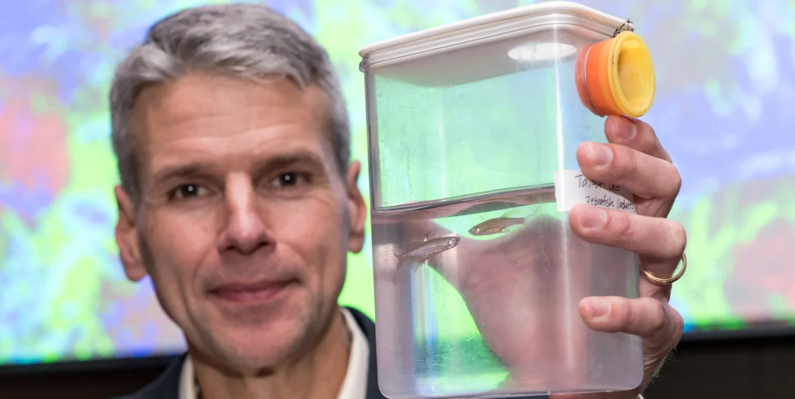
Will Talbot, PhD, knew the obvious question and decided to address it directly. “Why does the medical school employ someone who studies zebrafish?”
Will Talbot, PhD, knew the obvious question and decided to address it directly. “Why does the medical school employ someone who studies zebrafish?” Talbot asked the several hundred people assembled on the Stanford campus recently to celebrate the Discovery Innovation Awards, competitive seed grants that support early stage research in human biology.
A developmental biologist as well as a senior associate dean at the School of Medicine, Talbot explained that he studies the small striped creatures because, like humans, zebrafish are vertebrates that rely on myelin, a substance that is essential for a functioning nervous system. He said he can raise huge numbers of fish in his lab and peer straight into their nervous systems because the embryos and larvae are transparent.
Five years ago, Talbot said he wanted to study zebrafish that couldn’t make myelin to see if he could devise a strategy to reverse the defect. If he could restart myelin production in zebrafish, he knew it could have implications for treating people with multiple sclerosis, a disabling autoimmune disease caused by damage to the myelin sheath surrounding nerve cells.
At the time, Talbot said he couldn’t approach traditional funding sources for medical research — he didn’t have enough evidence to show them — but he needed resources. “How do you pay people in the lab?” he asked. “How do you feed the fish?”
Talbot applied for a Stanford Medicine Discovery Innovation Award, funded by philanthropists who believe that one discovery can have an exponential impact on human health. A committee of experts selected Talbot’s application and awarded him $95,000 to pursue a project with the mutant zebrafish.
His research revealed on molecular level why there was no myelin in the mutant fish. From there, he was able to reverse the defect. This line of research is so promising that other traditional funding organizations have pitched in.
“A few years ago,” he said, “they wouldn’t have even considered funding us.”
The Discovery Innovation Awards, which have distributed $6 million over the last four years, come with little red tape and few constraints. These grants have resulted in 119 research papers and more than $36 million in follow-on funding, according to Stanford Medical Center Development records.
At the campus event celebrating the most recent group of awardees, Talbot joined three other scientists who won the awards in previous years to talk about their research.
Geneticist Anne Brunet, PhD, described the methods that she developed to read each cell’s chromatin signature and understand the genetic mechanisms of aging and longevity. Computational biochemist Rhiju Das, PhD, discussed Eterna, an online game he created to crowdsource the design of molecular medicines. And David Schneider, PhD, talked about ways to increase resilience to infectious diseases.
Nobel Laureate Roger Kornberg, PhD, introduced the award winners and emphasized the importance of research that increases fundamental knowledge of human biology. “Science is hard and it’s unpredictable,” Kornberg said. “There’s no guarantee of success and discoveries are by their nature accidental. They depend on luck and serendipity and they require a long time.”
Kornberg was the first scientist to discover the three-dimensional structure of how DNA is converted into messenger RNA. This discovery showed how human cells — each of which contains a person’s entire genetic code — selectively read out sections and differentiate into all the types of cells necessary to sustain life.
“I started on the work while in my 30s,” he said. “When we succeeded, I was approaching 60.” He was awarded the Nobel Prize in Chemistry in 2006, six months shy of his 60th birthday.
Now, Kornberg said people often ask him what he believes are the most promising areas of research and what research should be funded.
“I answer with the obvious,” he said. “We cannot predict the future. All white papers can be replaced by a simple statement: support the brightest, most ambitious, most hard-working young scientists and they will invent the future.”
Jody Berger
Published on March 19, 2018

Memorial Celebration Honoring Ben Barres
Please join us for a memorial celebration and reception to honor the life and contributions of Ben Barres, professor of neurobiology, who passed away on December 27, 2017.
Date: Monday, April 2
Program: 1:00 pm
Reception: 2:45 pm
Location: McCaw Hall, Arrillaga Alumni Center, Stanford
Please join us for a memorial celebration and reception to honor the life and contributions of Ben Barres, professor of neurobiology, who passed away on December 27, 2017. The event is scheduled for 1:00 pm on April 2 at the Arrillaga Alumni Center and is open to members of the Stanford University community. If planning to attend, RSVP by March 21.

James Byers recipient of the Weintraub Award
Congratulations to James Byers in Daniel Jarosz lab for having been selected for a Harold M. Weintraub Graduate Student Award to recognize outstanding achievement in Graduate Studies.
It gives us great pleasure to inform you that James Byers in Daniel Jarosz lab has been selected for a Harold M. Weintraub Graduate Student Award to recognize outstanding achievement in Graduate Studies. This is a very prestigious award for a graduate student, honoring the late Hal Weintraub and given out by the Fred Hutchinson Cancer Research Center.

Neuroscience awards named in honor of Ben Barres
The five-year awards from the Chan Zuckerberg Initiative will help fuel research into the biology of neurodegenerative diseases. The awards honor a Stanford neuroscientist who died in December.
The Chan Zuckerberg Initiative, a Palo Alto-based philanthropic organization, has launched a major research effort to inject fresh energy, ideas and talent into understanding the basic biology of neurodegenerative conditions such as Alzheimer’s disease, Parkinson’s disease and amyotrophic lateral sclerosis, the organization announced Feb. 20.
The research will be funded through two programs, including one that will support early career investigators willing to pursue bold, innovative ideas. These five-year awards, known as the CZI Ben Barres Early Career Acceleration Awards, are named in honor of Ben Barres, PhD, a distinguished Stanford neuroscientist who died in December at the age of 63.
CZI will also fund a series of collaborative science awards — three-year grants for small, interdisciplinary groups of scientists, clinicians and engineers working together on innovative high-risk, high-impact projects in basic science.
Both grant programs are part of CZI’s new Neurodegeneration Challenge Network, which aims to fill in the gaps in the still-limited understanding of the basic cellular and molecular mechanisms behind these devastating illnesses.
Cori Bargmann, PhD, president of Chan Zuckerberg Science, said the group chose to name the young investigator awards in Barres’ honor because he was “a spiritual guide” for the work. An advocate for basic science and for the mentorship of young researchers, Barres had been an adviser to CZI since its inception in 2015 and had a hand in helping craft the awards program, Bargmann said.
‘Exceptional scientist and human being’
“Ben was a truly exceptional scientist and human being. He exemplified the values of the Chan Zuckerberg Initiative, especially our work in neurodegenerative disease. His commitment to collaboration between basic science and medicine, his creative work in neurodegeneration, and his advocacy for women, underrepresented groups and young scientists inspire us all,” Bargmann said.
Barres made significant discoveries about the role of glial cells, the under-recognized cells that comprise the majority of brain cells, and in doing so revolutionized the field of neuroscience. A professor of neurobiology, of developmental biology and of neurology, he was widely praised for the passion he brought to his work.
Barres was particularly known for his dedication to his trainees and was a champion for basic science, helping establish the Master of Science in Medicine program at Stanford to teach PhD students about human biology and disease and thus prepare them to turn new discoveries into clinically useful treatments.
“Ben was a selfless and steadfast champion of young researchers. As his colleague and friend, I am moved that these awards will commemorate him and continue his legacy of celebrating and supporting early-career scientists,” said neuroscientist Marc Tessier-Lavigne, PhD, president of Stanford University.
Lloyd Minor, MD, dean of the School of Medicine, said the awards “are an inspired way to honor the memory of Ben, a remarkable person and a beloved mentor who embodied the spirit of the awards in his brilliance, creativity and passion for neuroscience.”
The CZI Ben Barres Early Career Acceleration Awards are open to scientists from throughout the world working in a variety of disciplines. The awards are open to MDs and PhDs who are new to the field of neurodegeneration. Awardees will receive $500,000 a year for five years, for a total of $2.5 million. The collaborative science awards will provide recipients with $350,000 a year for three years, or a total of $1.05 million.
CZI was founded by Silicon Valley couple Priscilla Chan, MD, and Mark Zuckerberg, chairman and CEO of Facebook, to advance science, education and social justice. One of CZI’s first initiatives was the creation of the Chan Zuckerberg Biohub, an independent nonprofit research center supported by $600 million over a 10-year period. The center brings together physicians, scientists and engineers at Stanford, UC-San Francisco and UC-Berkeley to engage in innovative scientific exploration and invent new tools to advance discoveries.
Information about the awards is available online.

Dynamic DNA dance identified with new CRISPR/Cas9-based labeling
DNA twitches during transcription to bring distant regions in contact and enhance gene expression, according to Stanford researchers who devised a new way to label individual, nonrepetitive DNA sequences.
DNA flails about during transcription like a strand of spaghetti being sucked through pursed lips, School of Medicine researchers have found. Like the resulting out-of-control flying globs of sauce, the surprising discovery flies in the face of conventional wisdom, which posits that static loops of DNA are required to bring together distant regions that enhance and promote gene expression.
A new DNA labeling technique, which can precisely tag any individual stretch of DNA with fluorescent molecules to track their three-dimensional locations and movements, revealed this genetic dance. The technique, which the researchers have termed CARGO, for chimeric array of gRNA oligo, is a variation of the CRISPR/Cas9 gene-editing tool, and it promises to revolutionize the study of genome dynamics.
‘Entirely unexpected and surprising’
“We’ve found that, as the polymerase plows across the DNA, it provides a source of molecular agitation that increases mobility within a local chromosome domain and can repeatedly bring distant regions of the genome together,” said Joanna Wysocka, PhD, professor of developmental biology and of chemical and systems biology. “This was entirely unexpected and surprising, and directly counters the prevailing beliefs about transcription. It’s just one example of what we and others can now learn by using CARGO to label specific DNA regions.”
A paper describing the research was published Jan. 25 in Science. Wysocka is the senior author, and graduate student Bo Gu is the lead author.
CRISPR is most commonly used to seek out and replace specific DNA sequences in the genome with other DNA sequences. To do so, an enzyme called Cas9 uses a short RNA sequence to guide the DNA sequences to the correct spot in the genome.
A variation of the technique that was developed by other researchers instead uses guide RNAs and the CRISPR system with a catalytically inactive form of Cas9 to recognize and label specific stretches of DNA with fluorescent molecules. But that works best on highly repetitive regions where a single guide RNA can marshal the critical mass of fluorescent tags necessary to generate enough light to be seen through a microscope.
Wysocka, Gu and another co-author of the study, Tomasz Swigut, PhD, devised a way to introduce an array of many different guide RNAs into a cell to precisely recognize nonrepetitive, unique stretches of DNA and label them with multiple fluorescent tags so they can be easily visualized under a microscope.
‘CARGO solves the delivery problem’
“All the most interesting stuff in the genome is present as single copies,” Wysocka said. “People have been trying unsuccessfully to label single regions, or loci, for some time. But CARGO solves the delivery problem. Now we can label any region, or locus, that we want by using many different guide RNAs to blanket the DNA so we can see it clearly.” She and Gu emphasize that the CARGO technique will be useful to researchers pursuing many different questions about the genome or gene expression.
This was entirely unexpected and surprising, and directly counters the prevailing beliefs about transcription.
Already it’s opened their eyes about the process of transcription, which is often stimulated when distant enhancer regions are brought into close proximity with other DNA regions called promoters.
“We found that any locus we looked at moved about four times faster in its active state, when nearby genes are being transcribed into RNA,” Wysocka said. “We propose that this enhanced movement, or diffusion, is likely to bring distant regions of the DNA together and further promote transcription.”
Other Stanford authors of the study are graduate students Andrew Spencley and Mingyu Chung; former research technician Matthew Bauer, and professor of chemical and systems biology Tobias Meyer, PhD.
Gu, Swigut and Wysocka have filed a U.S. provisional patent application relating to the CARGO methodology.
The research was supported by the National Institutes of Health (grant GM112720), the Howard Hughes Medical Institute and a Stanford graduate fellowship.
Stanford’s departments of Developmental Biology and of Chemical and Systems Biology also supported the work.
By Krista Conger
Krista Conger is a science writer for the medical school's Office of Communication & Public Affairs. Email her at kristac@stanford.edu.

Irving Weissman honored by several organizations
Irving Weissman, MD, professor of pathology and of developmental biology, received several awards and an honorary doctorate in 2017.
Irving Weissman, MD, professor of pathology and of developmental biology, received several awards and an honorary doctorate in 2017.
He received the 2017 Donald Metcalf Award from the International Society for Experimental Hematology. The award recognizes distinguished society members who have made outstanding contributions to the field of experimental hematology.
Weissman, who holds the Virginia and D.K. Ludwig Professorship for Clinical Innovation in Cancer Research, also received a 2017 National Cancer Institute Outstanding Investigator Award, which recognizes accomplished leaders in cancer research and provides up to $600,000 a year for seven years. He plans to use the award to investigate whether mutations accumulate in a central nervous system stem cell clone that becomes a brain cancer stem cell.
He was awarded the 2017 Karl Landsteiner Memorial Award and Lectureship by the American Association of Blood Banking. The honor recognizes a scientist who has an international reputation in transfusion medicine or cellular therapies. Weissman was recognized for his pioneering role identifying and isolating the first hematopoietic stem cells in mice and humans.
He was awarded the Helmholtz International Fellow Award from the Helmholtz Association. The prize includes 20,000 euros (about $24,000) and an invitation to conduct research at one of the Helmholtz Centres. The Helmholtz Association of German Research Centres, funded by the German government, conducts research related to challenges facing society in several fields, including health.
In addition, he received an honorary doctorate from the Faculty of Medicine at the University of Turku in Finland.
Neuroscientist Ben Barres, who identified crucial roles of glial cells, dies at 63
The Stanford neuroscientist’s research focused on the cells in the brain that aren’t nerve cells. Collectively called glia, these “other” cells play a central role in sculpting and maintaining the brain’s wiring diagram.
The Stanford neuroscientist’s research focused on the cells in the brain that aren’t nerve cells. Collectively called glia, these “other” cells play a central role in sculpting and maintaining the brain’s wiring diagram.
Acclaimed Stanford neuroscientist Ben Barres, MD, PhD, died on Dec. 27, 20 months after being diagnosed with pancreatic cancer. He was 63.
Barres’ path-breaking discoveries of the crucial roles played by glial cells — the unsung majority of brain cells, which aren’t nerve cells — revolutionized the field of neuroscience.
Barres was incontestably visionary yet, ironically, face-blind — he suffered from prosopagnosia, an inability to distinguish faces, and relied on voices or visual cues such as hats and hairstyles to identify even people he knew well. And there were many of them.
A professor of neurobiology, of developmental biology and of neurology, Barres was widely praised as a stellar and passionate scientist whose methodologic rigor was matched only by his energy and enthusiasm. He was devoted to his scholarly pursuits and to his trainees, advocating unrelentingly on their behalf. He especially championed the cause of women in academia, with whom he empathized; he was transgender.
“Ben was a remarkable person. He will be remembered as a brilliant scientist who transformed our understanding of glial cells and as a tireless advocate who promoted equity and diversity at every turn,” said Marc Tessier-Lavigne, PhD, president of Stanford University. “He was also a beloved mentor to students and trainees, a dear friend to many in our community and a champion for the fundamental dignity of us all.” (Read Tessier-Lavigne's tribute to Barres.)
Added Lloyd Minor, MD, dean of the School of Medicine, “Through courage and determination, Ben not only changed the course of neuroscience, he touched many lives. He was an inspiration, and I, like so many others, am a better person for having known him.”
Nine of every 10 brain cells
Barres’ research focused on the nine of every 10 cells in the human brain that aren’t nerve cells, or neurons. They’re called glial cells or, collectively, glia.
“Ben pioneered the idea that glia play a central role in sculpting the wiring diagram of our brain and are integral for maintaining circuit function throughout our lives,” said Thomas Clandinin, PhD, professor of neurobiology, who assumed the role of departmental chair in April 2016 when Barres, who had held the position from 2008 until then, was first diagnosed with pancreatic cancer. “People had thought glia were mere passive participants in maintaining neural function. Ben’s own work and that of his trainees transformed this view entirely.”
When Barres first began studying them, glia, whose name comes from the Greek word for glue, were thought to be not much more than packing peanuts, supplying positional stability and various nutrients to the brain’s much more talented neurons.
But Barres and the numerous trainees who cycled through his lab showed otherwise.
Glial cells, they proved, are critical to sustaining the overall architecture of the brain’s constellation of synapses, through which neurons pass signals to one another. Recent evidence from Barres’ lab indicates that glia gone wrong may be to blame for many of the neurodegenerative disorders that vex humanity.
“Ben placed a big career bet on the possibility that there was gold in glia,” said neurobiology professor William Newsome, PhD, the Vincent V. C. Woo Director of the Stanford Neurosciences Institute. “And he started by solving a big problem: No one had been able to grow glial cells in isolation.”
Burning the midnight oil
Intent on determining exactly how glia influence brain function and dysfunction, Barres typically worked until midnight or later throughout his career. Early on, he generated tools that allowed each of the three distinct types of glial cells to be purified and cultured in a way that retained all of their functionality, so they could be studied in a dish with a previously unobtainable acuity. Rather than jealously guard his methods and reagents, Barres took pains to make them widely available to others just as, later on, he did with the voluminous data his lab was able to generate with them.
“He had a selfless, outward-looking focus,” Clandinin said. “I’ve gone a lot of places in the months since Ben was diagnosed, and I haven’t gone anywhere yet where someone hasn’t come up to me and asked me about how Ben was doing. Every one of them has a story about how he helped them in their career.”
In doing so, Barres seeded an entire field of scientists studying glia, said Andrew Huberman, PhD, an associate professor of neurobiology at Stanford who was Barres’ postdoctoral advisee from 2005 through 2010.
“He didn’t have these normal territorial issues all of us have,” Huberman said. “He always gave more than he took. If ever there was an example of a purpose-driven life, it’s Ben. His passion was for science. His obsession was glia. His mission was to bring equality to how people are treated and promoted in science.”
Born Sept. 13, 1954, Barres grew up in West Orange, New Jersey, one of four children in a not well-to-do family. He got his first taste of science in the West Orange Public Library, developed an affinity for microscopes and chemistry sets, and became a high school math star. Attending the Massachusetts Institute of Technology on a scholarship, he earned a bachelor’s degree in life science there in 1976 and headed to medical school at Dartmouth, where he obtained an MD in 1979.
Motivated by a mystery
During his subsequent internship and residency in clinical neurology at Cornell, Barres grew increasingly frustrated at physicians’ inability to provide cures or even to understand the causes of neuronal degeneration. He was struck by the observation, in pathologists’ specimens of degenerating brain tissue, of irregular-appearing glial cells’ ubiquitous presence near the lesions.
Bent on finding out why, Barres changed course. He returned to academia, enrolling in a graduate program in Harvard Medical School’s neuroscience program in 1983, and published several research papers by the time he received his PhD in neurobiology in 1990. Then he embarked on a postdoctoral fellowship in the lab of Martin Raff, MD, a professor of biology at University College London who was using immunological techniques to tease apart the three classes of glial cells.
Working under Raff, Barres pushed forward and unearthed new insights concerning the best-known glial class: oligodendrocytes, cells stuffed with a fatty substance called myelin. These fat-filled cells were already understood to wrap themselves around neurons’ lengthy projections, a process called myelination, providing electrical insulation and vastly increasing the transmission speed and reliability of neuronal impulses. Barres showed, among other things, that electrical activity in neurons was necessary for neurons’ myelination.
Barres would routinely work in the lab until 2 or 3 a.m., said Raff. “He slept on the floor of my small office. Every morning when I arrived and opened the door, it would whack him in the head — he eventually learned to sleep facing the opposite direction.”
Arriving at Stanford
In 1993, Barres moved from University College London to an assistant professorship in Stanford’s Department of Neurobiology. He was promoted to associate professor of neurobiology and of developmental biology in 1998, and to a full professorship in 2001. In 2008, he became chair of neurobiology. From 2005 on, he was the director of the Masters of Science in Medicine Degree Programfor PhD students, which he had created.
At Stanford, Barres turned his attention to a second class of glial cells known as astrocytes. These are the most common cells in the human brain, outnumbering neurons by a factor of four or so. Before Barres began focusing on them, nobody really had understood what astrocytes do for a living. With his colleagues, he discovered that they are crucial to the physical formation of synapses, as well as to those synapses’ functional activation. He and his colleagues also discovered that astrocytes cooperate with microglia — a third glial-cell type that’s become the object of much recent attention in Barres’ lab — in pruning away excess synapses during fetal and neonatal development, in essence preserving brain circuitry that’s proven itself to perform legitimate activities and clearing out the dead wood.
Beth Stevens, PhD, then a postdoctoral scholar in Barres’ lab, led a 2007 study showing that the cooperation of astrocytes and microglia in synaptic pruning involves the coordinated secretion of molecules previously thought to be exclusive to the body’s immune system. Stevens continues to focus on this phenomenon as an associate professor of neurology at Harvard Medical School.
“When I left Stanford for my new job,” she said, “Ben told me, ‘Take this work with you to your new lab, Beth. Nobody can do it better than you.’ Mentors aren’t always so generous about ceding areas of research initiated in their lab to trainees headed elsewhere. But Ben was a very special person. Not only was he an incredible scientist, but he also cared deeply about other people, especially his trainees. We were his kids.”
Never losing sight of goal
Barres never lost sight of his original goal: to figure out the molecular and cellular causes of the brain tissue degeneration seen in Alzheimer’s, Parkinson’s and Huntington’s diseases; multiple sclerosis; amyotrophic lateral sclerosis, or Lou Gehrig’s disease; and glaucoma, an optic-nerve degenerative disease. Research in Barres’ lab has strongly implicated inflamed or “reactive” astrocytes and microglia as drivers in all of these neurodegenerative disorders — most recently, in a 2017 Nature paper describing how certain reactive astrocytes secrete something that kills stressed or injured neurons.
In an interview about this study, Barres described these findings as “the most important discovery my lab has ever made.”
“Wherever we look in degenerating cortical tissue, we find reactive astrocytes,” said the study’s lead author, Shane Liddelow, PhD, a postdoctoral scholar in Barres’ lab. “And now we’ve learned that a subset of these reactive astrocytes not only fail to execute their synapse-building and -pruning tasks but also secrete a factor, or combination of them, that’s toxic to damaged neurons, and that these astrocytes become malevolent only when stimulated by yet other factors secreted by microglia that are themselves in an inflammatory state.”
Postdoctoral scholar Mariko Bennett, PhD, who will receive a medical degree in June, identified those microglia-derived factors, and graduate student Kevin Guttenplan is working on identifying and characterizing the astrocyte-generated toxin. In 2011, Barres co-founded a biotechnology company, Annexon Biosciences, to translate these findings into drugs that could someday succeed in retarding or preventing the progression of neurodegenerative disorders.
‘Just so I could work with him’
Liddelow hadn’t initially intended to study glia. In 2010, he was a graduate student in Australia, focused on another research area. “I met Ben at a meeting, and we hit it off. I switched fields in a heartbeat, just so I could work with him.”
Liddelow sat next to Barres during the meeting. “He was happily handing me one after another peanut butter and jelly sandwich,” Liddelow recalled. “I didn’t like peanut butter. But I think I ate three sandwiches. I just wanted him to like me.”
Barres was an outspoken champion of marginalized minorities in academia and society, not infrequently digressing for a few minutes during his scientific talks to point out the differences he’d personally experienced in how other scientists treated him when they perceived him as a woman versus as a man.
Barres spent his last days and final hours making sure that the letters of recommendation he had written for others were ready. “In what time remains to me that will be my highest priority,” he assured trainees in a letter he sent to them in early November.
Over the course of his career, Barres’ published 167 peer-reviewed papers, organized and chaired numerous meetings, won many awards and served on the editorial boards of Science, Neuron, the Journal of Neuroscience, the Journal of Cell Biology, Glia, Current Biology and more. He was elected to membership in the American Association for the Advancement of Science, the American Academy of Arts and Sciences, the National Academy of Sciences and the National Academy of Medicine.
“If you took the Barres lab out of the field of glial studies, there would be no field,” Raff said.
Much of that field was in attendance for a celebratory symposium/reunion held in Barres’ honor at Stanford on Jan. 12, 2017. “It was like a giant lab meeting,” said Stevens, one of the organizers. “Everybody came except for a handful who couldn’t make it for logistical reasons. We’re really a tightly knit family. And Ben was the nucleus that kept us all together.”
By Bruce Goldman
Bruce Goldman is a science writer for the medical school’s Office of Communication & Public Affairs. Email him at goldmanb@stanford.edu.

Beautiful Piles of Bones: An Interview with 2017 Genetics Society of America Medal Recipient David M. Kingsley
The Genetics Society of America Medal is awarded to an individual for outstanding contributions to the field of genetics in the last 15 years.
Abstract
The Genetics Society of America Medal is awarded to an individual for outstanding contributions to the field of genetics in the last 15 years. Recipients of the GSA Medal are recognized for elegant and highly meaningful contributions to modern genetics, exemplifying the ingenuity of GSA membership. The 2017 recipient is David M. Kingsley, whose work in mouse, sticklebacks, and humans has shifted paradigms about how vertebrates evolve. Kingsley first fell in love with genetics in graduate school, where he worked on receptor mediated endocytosis with Monty Krieger. In his postdoctoral training he was able to unite genetics with his first scientific love: vertebrate morphology. He joined the group of Neal Copeland and Nancy Jenkins, where he led efforts to map the classical mouse skeletal mutation short ear. Convinced that experimental genetics had a unique power to reveal the inner workings of evolution, Kingsley then established the stickleback fish as an extraordinarily productive model of quantitative trait evolution in wild species. He and his colleagues revealed many important insights, including the discoveries that major morphological differences can map to key loci with large effects, that regulatory changes in essential developmental control genes have produced advantageous new traits, and that nature has selected the same genes over and over again to drive the stickleback’s skeletal evolution. Recently, Kingsley’s group has been using these lessons to reveal more about how our own species evolved.
This is an abridged version of the interview. The full interview is available on the Genes to Genomes blog, at genestogenomes.org/kingsley/.
What inspired you to become a scientist?
My dad died of cancer when he was 34. As a little kid I was aware that you don’t know how long you have left, and I grew up wanting to make sure I spent the time I have doing something interesting and important. I thought that tackling age-old mysteries about life’s origin and mechanisms was a good way to spend my life.
What did you learn from your first mentors?
I was a kid who loved dinosaurs and skeletons. That interest was nurtured by a great high school teacher, Jack Koch at Roosevelt High School in Des Moines, Iowa. In his advanced biology class we memorized the names of every bone and muscle in the cat and human skeleton. A lot of people hated it, but I loved it because you could see so much about the function and lifestyle of the organisms from the size and shapes and patterns of bones.
In graduate school I fell in love with the power of genetics. I had a set of teachers at MIT, including David Botstein and Monty Krieger, who helped me learn that with genetics you didn’t have to assume anything about the answer to your question. You didn’t have guess that you were looking for a particular type of molecule or anything like that. Genetics was an algorithm that would take you to the key components controlling a biological system no matter what they were.
Why did you choose to work on the short ear gene?
Vertebrate genetics takes a long time, so you should pick your problem carefully. I didn’t want to pick something that was better studied in bacteria, yeast, or powerful invertebrate systems. The skeleton was perfect; it’s the defining feature of vertebrates. It also plays such an important role in animals’ external appearance that many classic mutants had already been picked up in simple morphological screens.
After World War II there had been a lot of interest in the effects of radiation on the mammalian germline, and there were two big mouse forward mutation experiments in the UK and US. They both used a test strain carrying seven homozygous recessive mutations with visible phenotypes. These were six pigment mutations and short ear. Millions of wild type mice were mutagenized and crossed with the test strain to measure the rate that new alleles were recovered at any of the seven loci. As a result, there were lots of newly induced mutations, including a whole set of deficiency chromosomes that took out both short ear and one of the closely linked pigmentation loci. We essentially had the equivalent of a Drosophila genetics playground for this particular region of the mouse genome!
What did you learn from the short ear project?
It took ∼5 years to do the chromosome walk in the region, and I was already an assistant professor by the time we eventually isolated the gene for this classic skeletal trait. But it was incredibly gratifying. The gene controlling skeletal morphology encoded a secreted signal already named a “bone morphogenetic protein” (BMP). It had been named by biochemists who found that if you took an adult bone and ground it into powder and injected it under the skin of an animal, there was some magic ingredient that could generate a brand new bone at the site of implantation.
The short ear mice provided the first genetic evidence that BMPs were the endogenous signals that vertebrates were using to set the form and pattern of skeletal structures. The large collection of short ear mutations later helped us to identify a whole series of modular, remarkably specific enhancers controlling different aspects of skeletal morphology. For someone originally interested in those beautiful piles of bones, to be able to break down their shapes into the expression patterns of secreted signaling molecules was an incredibly satisfying answer.
Why did you choose sticklebacks?
If you can find a way to turn old biological problems into genetics problems, then you can often find the answers to even intractable questions. A brave postdoc, Katie Peichel, and I spent a really fun summer in 1998 figuring out how to turn classic evolutionary questions into a genetics problem. We wanted to identify the number and type of genes and mutations that control species differences in nature. We went around talking to biologists, reading all kinds of books, looking for very young species with recently evolved and dramatic skeletal differences that could still be crossed in the laboratory. Somewhere in the middle of that summer I found a great book chapter by Mike Bell of Stonybrook University talking about all the cool skeletal traits that had evolved in sticklebacks after the end of the last Ice Age. There was a remarkable previous literature on stickleback morphology, ecology, and behavior in new freshwater streams and lakes. And new forms had evolved not just once but thousands of times. It was like nature had set off a replicate series of evolution experiments 10,000 years ago, producing new forms over and over again.
What did you learn about the repeatability of evolution?
I had a debate with a fellow faculty member when I started the project because he thought the project was not worth doing. It would just turn out to be postage stamp collecting, and there wouldn’t be any generality.
At the time, we didn’t have evidence one way or another. But my best reply was: how do you know? That was the great thing about genetics—it would tell you the answer no matter what the answer is. We started crossing these fish with huge skeletal differences. And if you compared the results from crosses done in different lakes, it turned out the very same chromosome regions were being used over and over again in different populations.
We’ve subsequently taken lots of traits down to genes and molecules. We’ve found that the key signals and transcription factors that developmental biologists have been studying for years turn out to be the same molecules that nature is using to redesign anatomical features. For example, although we didn’t set out to test any particular candidate genes, the genetic data showed us that some of those stickleback skeletal traits are controlled by the same kinds of bone morphogenetic proteins that we found in mouse.
How does the stickleback work connect with your studies of human evolution?
We’re interested in why particular genes are reused throughout evolution, and we’re also interested in applying the patterns we’ve found in sticklebacks to the evolution of ourselves. We’ve found that classic traits in people, like blond hair color, or height, are evolving in humans using the same types of key control genes and regulatory mutations we have found in fish. And, unlike rare genetic diseases, there are derived alleles at these human loci where a large fraction of the population carry the selected version. In some cases, the selected alleles may actually alter susceptibility to late onset diseases like skin cancer or arthritis. It’s not a huge effect, maybe 1.3- to 1.8-fold. But when an allele slightly increases risk of a disease and is carried by a few billion people through selection, then suddenly you find an awful lot of the burden of a common human disease is controlled by our own evolutionary history. We’re now going back and forth between humans and the patterns we see in fish. We thought it might take us 50 years to get enough examples to pull out general principles, but it turned out to be much faster that.
- Copyright © 2017 by the Genetics Society of America

Second ‘don’t eat me’ signal found on cancer cells
CD47 is an important inhibitor of cancer-killing immune cells called macrophages. Now Stanford researchers have identified another, similar way to activate macrophages to destroy cancer cells.
CD47 is an important inhibitor of cancer-killing immune cells called macrophages. Now Stanford researchers have identified another, similar way to activate macrophages to destroy cancer cells.
NOV 27 2017
Irving Weissman and his collaborators have found a second pathway that could be used in efforts to boost the body's ability to kill cancer cells.
Norbert von der Groeben
A second biological pathway that signals immune cells not to engulf and kill cancer cells has been identified by researchers at the Stanford University School of Medicine.
An antibody that blocks the “don’t eat me” signal has shown promise as a cancer treatment in animal models and is currently in clinical trials. Combining that antibody, known as anti-CD47, with another that blocks this newly discovered pathway could further enhance the ability of the immune system to eradicate many types of cancers, the researchers believe.
“The development of cancer cells triggers the generation of SOS molecules recognized by the body’s scavenger cells, called macrophages,” said Irving Weissman, MD, the director of Stanford’s Institute for Stem Cell Biology and Regenerative Medicine, and also of its Ludwig Cancer Center. “However, aggressive cancers express a ‘don’t eat me’ signal in the form of CD47 on their surfaces. Now we’ve identified a second ‘don’t eat me’ signal and its complementary receptor on macrophages. We’ve also shown that we can overcome this signal with specific antibodies and restore the ability of macrophages to kill the cancer cells.”
A paper describing the findings was published online Nov. 27 in Nature Immunology. Weissman, a professor of pathology and of developmental biology, shares senior authorship of the study with former postdoctoral scholar Roy Maute, PhD, who is now head of biology at Ab Initio Biotherapeutics Inc. Graduate student Amira Barkal shares lead authorship with former graduate student Kipp Weiskopf, MD, PhD, who is now a resident at Brigham and Women’s Hospital.
“Simultaneously blocking both these pathways in mice resulted in the infiltration of the tumor with many types of immune cells and significantly promoted tumor clearance, resulting in smaller tumors overall,” Barkal said. “We are excited about the possibility of a double- or perhaps even triple-pronged therapy in humans in which we combine multiple blockades to cancer growth.”
Importance of macrophages
Macrophages are large white blood cells found in nearly all the body’s tissues. As part of what’s known as the innate immune system, they engulf and kill foreign invaders like bacteria or viruses. They also destroy dead and dying cells and, in some cases, cancer cells whose internal development cues have gone haywire.
The “don’t eat me” signal was identified in Weissman’s laboratory in 2009. His team found that nearly all cancer cells express high levels of a molecule called CD47 on their surfaces. They showed that CD47 binds to a protein called SIRPalpha on the surface of macrophages, inhibiting their ability to kill the cancer cells.
Animal studies showed that treatment with an anti-CD47 antibody vastly improved the ability of macrophages to kill cancer cells and even led to some cures in mouse models of cancer. Phase-1 clinical trials are currently underway at Stanford and in the United Kingdom to test the safety and efficacy of the treatment in humans with a variety of blood and solid tumors.
The newly discovered binding interaction used by cancer cells to evade macrophages capitalizes on a protein structure on the cancer cells’ surface called the major histocompatibility complex class 1, or MHC class 1. Human tumors that have high levels of MHC class 1 on their surfaces are more resistant to anti-CD47 treatment than are those with lower levels of the complex, the researchers found.
Component of adaptive immunity
MHC class 1 is an important component of adaptive immunity, the second major arm of the immune system, which relies on immune cells called T cells and B cells to nimbly and specifically respond to foreign invaders and cell damage. Most cells of the body express MHC class 1 on their surfaces as a way to indiscriminately display bits of many proteins found within the cell — a kind of random sampling of a cell’s innards that provides a window into its health and function. If the protein bits, called peptides, displayed by the MHC are abnormal, a T cell destroys the cell. Although the relationship between MHC class 1 and T cells has been well-established, it’s been unclear whether and how the complex interacts with macrophages.
These findings help us understand the many ways cancer cells can evade macrophages, and how we might block these escape pathways.
Barkal and her colleagues found that a protein called LILRB1 on the surface of macrophages binds to a portion of MHC class 1 on cancer cells that is widely shared across individuals. This binding inhibits the ability of macrophages to engulf and kill the cancer cells, both when growing in a laboratory dish and in mice with human tumors, the researchers found. Inhibiting both the CD47-mediated pathway and the LILRB1 pathway significantly slowed tumor growth in mice.
Understanding the balance between adaptive and innate immunity is important in cancer immunotherapy. For example, it’s not uncommon for human cancer cells to reduce the levels of MHC class 1 on their surfaces to escape destruction by T cells. People with these types of tumors may be poor candidates for cancer immunotherapies meant to stimulate T cell activity against the cancer. But these cells may then be particularly vulnerable to anti-CD47 treatment, the researchers believe. Conversely, cancer cells with robust MHC class 1 on their surfaces may be less susceptible to anti-CD47.
“In some cancers, MHC class 1 expression, for a variety of reasons, is not reduced,” Weissman said, “and this helps the cancer cells escape from macrophages. These findings help us understand the many ways cancer cells can evade macrophages, and how we might block these escape pathways.”
“The fact that there are at least two redundant mechanisms to modulate macrophage activity is a testament to how critically important it is to tightly control our immune responses,” Barkal said. “It’s possible that future studies will identify even more of these pathways, which will give us additional targets for cancer immunotherapy.”
The research was supported by the National Institutes of Health (grants R01CA086017, R01GM100315, HL120824 and GM07365), the D.K. Ludwig Fund for Cancer Research, the Cancer Research Institute, the Human Frontier Science Program Organization, the University of Wisconsin Medical Scientists Training Program, the National Research Award, the Paul and Daisy Soros Fellowship for New Americans, the Stanford Medical Scientist Training Program and anonymous donors.
Other Stanford authors are technician Kevin Kao; former graduate student Sydney Gordon, PhD; postdoctoral scholar Benyamin Rosental, PhD; graduate students Ying Yiu, Benson George, Jonathan Tsai and James Chen; research associate Maxim Markovic; former medical fellow Nan Ring, MD; former research assistants Kelly McKenna and Po Yi Ho; and former undergraduate student Robin Cheng.
Weissman, Maute and Weiskopf are co-inventors on a patent related to the current work and own stock in FortySeven, Inc., which is pursuing clinical approval of the anti-CD47 antibody. Weissman, Maute, Weiskopf and James Chen are stockholders or consultants or employees of the company.
Stanford’s Department of Pathology also supported the work.
By Krista Conger
Krista Conger is a science writer for the medical school's Office of Communication & Public Affairs. Email her at kristac@stanford.edu.

SDRC becomes a NIH diabetes research center
The Stanford Diabetes Research Center (SDRC) announced that it has received a major program grant from the National Institutes of Health (NIH).
The Stanford Diabetes Research Center (SDRC) announced that it has received a major program grant from the National Institutes of Health (NIH). Stanford is only one of a handful of U.S. institutions to be supported as a NIH ‘Diabetes Research Center’. This is the first time Stanford has garnered this prestigious center designation
The SDRC is comprised of over 90 distinguished members from multiple schools at Stanford, including faculty in the Schools of Medicine, Engineering, and Arts and Sciences. These members are united by a common interest in understanding, treating and curing diabetes, including type 1 and type 2 diabetes, and forms of diabetes linked to pancreatic cancer. To support this work, the SDRC provides Research Cores, a Pilot and Feasibility Grant Program, Enrichment Activities, and other functions to support diabetes-related research at Stanford. The SDRC leadership includes its Director, Dr. Seung Kim, Professor of Developmental Biology and, by courtesy, of Medicine (Endocrinology and Oncology Divisions), and the SDRC Associate Director, Dr. David Maahs, Professor of Pediatrics, and Chief of the Division of Pediatric Endocrinology.
“Stanford has a superb group of investigators dedicated to transformative diabetes research, whose efforts are coordinated and aligned by the SDRC,” stated Kim. “We are thrilled to have earned this important support from the NIH to foster the SDRC programs. We also view this as important recognition of the outstanding and enduring fundamental and clinical investigations at Stanford that focus on understanding and treating diabetes and its complications. Unfortunately, diabetes incidence in its major forms is increasing worldwide. So this support is timely.”
The program project (P30) award for 7.7 million dollars from the NIDDK will provide support over the next five years for key functions of the SDRC.

Genome analysis with near-complete privacy possible
Stanford researchers used cryptography to cloak irrelevant genetic information in individuals’ genomes while revealing disease-associated mutations. They say the technique could vastly improve patient privacy.
Stanford researchers used cryptography to cloak irrelevant genetic information in individuals’ genomes while revealing disease-associated mutations. They say the technique could vastly improve patient privacy.
It is now possible to scour complete human genomes for the presence of disease-associated genes without revealing any genetic information not directly associated with the inquiry, say Stanford University researchers.
This “genome cloaking” technique, devised by biologists, computer scientists and cryptographers at the university, ameliorates many concerns about genomic privacy and potential discrimination based on an individual’s genome sequence.
Using the technique, the researchers were able to identify the responsible gene mutations in groups of patients with four rare diseases; pinpoint the likely culprit of a genetic disease in a baby by comparing his DNA with that of his parents; and determine which out of hundreds of patients at two individual medical centers with similar symptoms also shared gene mutations. They did this all while keeping 97 percent or more of the participants’ unique genetic information completely hidden from anyone other than the individuals themselves.
“We now have the tools in hand to make certain that genomic discrimination doesn’t happen,” said Gill Bejerano, PhD, associate professor of developmental biology, of pediatrics and of computer science. “There are ways to simultaneously share and protect this information. Now we can perform powerful genetic analyses while also completely protecting our participants’ privacy.”
Bejerano shares senior authorship of the research, which was published Aug. 18 in Science, with Dan Boneh, PhD, professor of computer science and of electrical engineering. Graduate students Karthik Jagadeesh and David Wu share lead authorship of the study.
Applying cryptography techniques
The researchers hope that routine implementation of their technique will help individuals overcome any qualms about privacy that may keep them from sharing their genome sequences. In particular, people may be concerned that DNA sequences or genetic variants currently unassociated with diseases may in the future be linked with as-yet-unidentified increases in risk.
Dan Boneh
“These are techniques that the cryptography community has been developing for some time,” said Boneh, who is the Rajeev Motwani Professor in the School of Engineering. “Now we are applying them to biology. Basically, if you have 1 million people with genomic data they would like to keep private, this approach lets researchers analyze the data in aggregate and only report on findings that are pertinent. An individual might have dozens of anomalous genes, but the researchers and clinicians will only learn about the genes relevant to the study, and nothing else.”
When the human genome was fully sequenced in 2001, it was hailed as a remarkable achievement. For the first time, the 3 billion nucleotides that encode the approximately 20,000 genes that keep our bodies running smoothly were tidily listed as a string of letters. But every human has many variations from the published, consensus sequence. These individual differences are what make us unique, but they can also confer increased risk of genetic diseases.
More than 7,000 diseases are caused by variations in the sequence of a single gene. But in order to determine which variations cause the condition, it has been necessary until now to compare the genetic sequences of hundreds or thousands of individuals with and without the disease, letter by letter. Geneticists (or their computer software) then make a list of all the differences and identify which are found primarily in people with the disease under study but rarely in any unaffected people. Those variations are then considered to be prime disease-causing suspects.
“There is a general conception that we can only find meaningful differences by surveying the entire genome,” said Bejerano. “But these meaningful differences make up only a very tiny proportion of our DNA. There are now amazing tools in computer science and cryptography that allow researchers to pinpoint only these differences while keeping the remainder of the genome completely private.”
In 2008, President George W. Bush signed the Genetic Information Nondiscrimination Act, which prohibits discrimination in matters of health insurance and employment based on an individual’s genetic information. But there are many other arenas in which such discrimination could potentially occur, including the purchase of life or disability insurance or applying for a loan.
Giving power to the individual
Jagadeesh and Wu worked together to adapt a cryptographic approach known as Yao’s protocol and cloud computing for use with human genomes. A key component of the technique is the involvement of the individual whose genome is to be studied. In particular, each individual encrypts their genome (with the help of a simple algorithm on their own computer or smart phone) into a linear series of values describing the presence or absence of the gene variants under study, without revealing any other information about their genetic sequence. The encrypted information is uploaded into the cloud and the researchers then use a secure, multi-party computation (a cryptographic technique that ensures the input data remain private) to conduct the analysis and reveal only those gene variants likely to be pertinent to the investigation.
We now have the tools in hand to make certain that genomic discrimination doesn’t happen.
“In this way, no person or computer, other than the individuals themselves, has access to the complete set of genetic information,” said Bejerano. In each case, the analysis was performed within seconds or minutes with moderate computing power. They hope to extend the technique to include diseases caused by combinations of multiple genetic variants or to handle tens of thousands of sequences such as those found in genome-wide association studies.
Ultimately the goal is to find the best way to both share the genetic information with researchers while also protecting each patient’s privacy in order to advance medical knowledge.
“Often people who have diseases, or those who know that a particular genetic disease runs in their family, are the most reluctant to share their genomic information because they know it could potentially be used against them in some way,” said Bejerano. “They are missing out on helping themselves and others by allowing researchers and clinicians to learn from their DNA sequences.”
Bejerano is a member of Bio-X, the Stanford Child Health Research Institute, the Stanford Cancer Institute and the Stanford Neurosciences Institute.
Another Stanford study co-author is graduate student Johannes Birgmeier.
The study was funded by Stanford University fellowship grants, the National Science Foundation, the Defense Advanced Research Projects Agency, the David and Lucile Packard Foundation, Microsoft and the Simons foundation.
Stanford’s departments of Developmental Biology, of Pediatrics and of Computer Science also supported the work.
- By Krista Conger
Krista Conger is a science writer for the medical school's Office of Communication & Public Affairs. Email her at kristac@stanford.edu.

In northern humans, evolution favored shorter bones — but with a painful trade-off
Early humans evolved to have shorter bones and an increased risk of osteoarthritis, a trade-off that may have helped them in colder climates, Stanford researchers say.
Jul 3 2017
David Kingsley
A single genetic change linked both to a reduction in human height and an increase in osteoarthritis risk might seem like it would quickly be kicked to the evolutionary curb. After all, how could it be an advantage to be both shorter and less mobile in the cutthroat competition for scarce resources and fickle mates? Darwin’s finches would be appalled.
Now, researchers at the Stanford University School of Medicine and at Harvard University have shown that, despite its association with the painful joint disease, this genetic variant has been repeatedly favored as early humans migrated out of Africa and into colder northern climates. At least half of Europeans and Asians harbor the gene variant, which is relatively rare in African populations.
“Because it’s been positively selected, this gene variant is present in billions of people,” said David Kingsley, PhD, professor of developmental biology at Stanford. “So even though it only increases each person’s risk by less than twofold, it’s likely responsible for millions of cases of arthritis around the globe. This study highlights the intersection between evolution and medicine in really interesting ways, and could help researchers learn more about the molecular causes of arthritis.”
A more compact body structure due to shorter bones could have helped our ancestors better withstand frostbite and reduce the risk of bone fracture from falling, the researchers speculate. These advantages in dealing with chilly temperatures and icy surfaces may have outweighed the threat of osteoarthritis, which usually occurs after prime reproductive age.
Cold may have selected for gene variant
“The gene we are studying shows strong signatures of positive selection in many human populations,” said Kingsley, who is also a Howard Hughes Medical Institute investigator and a member of Stanford Bio-X. “It’s possible that climbing around in cold environments was enough of a risk factor to select for a protective variant even if it brought along an increase likelihood of an age-related disease like arthritis, which typically doesn’t develop until late in life.”
A paper describing the research was published online July 3 in Nature Genetics. Kingsley is the senior author. Harvard graduate student Jiaxue Cao and former Stanford postdoctoral scholars Terence Capellini, PhD, and Hao Chen, PhD, share lead authorship. Capellini is now an associate professor of human evolutionary biology at Harvard, and Chen is an associate clinical scientist at Genentech Inc.
Many people think of osteoarthritis as a kind of wear-and-tear disease, but there’s clearly a genetic component at work here as well.
The researchers were studying a gene called GDF5 that Kingsley’s laboratory first linked to skeletal growth in the early 1990s. GDF5 is involved in bone growth and joint formation, and mutations in the coding portion of the gene have been shown to cause malformations in leg-bone structure in mice. In humans, GDF5 mutations are associated with shorter stature and joint problems; in particular, two nucleotide changes immediately upstream of the gene have been strongly associated with a 1.2- to 1.8-fold increase in the risk of osteoarthritis.
In the new study, the researchers were interested in learning more about how the DNA sequences surrounding GDF5 might affect the gene’s expression. Often, these noncoding sequences contain key regulatory regions known as promoters and enhancers. Capellini, Chen and Cao were able to identify a previously unknown enhancer region they termed GROW1, which is several thousand nucleotides downstream of GDF5.
When the researchers analyzed the sequence of GROW1 in the 1,000 Genomes Project database, which collects and compares sequences from many human populations around the globe, they identified a single nucleotide change that is highly prevalent in Europeans and Asians but that rarely occurs in Africans. When they introduced this nucleotide change into laboratory mice, they found that it decreased the activity of GDF5 in the growth plates of the long bones of fetal mice.
A common thread
Further research showed that this nucleotide change has been repeatedly favored during human evolution. Modern humans migrated from Africa between 50,000 and 100,000 years ago. But they weren’t the first to leave the continent. Neanderthals and Denisovans moved north into Europe and Asia about 600,000 years ago. Interestingly, the researchers found that the same GROW1 variant was found in the DNA of both ancient and modern humans in Europe and Asia.
However, there’s a dark side to this stocky, hardy body type: The GDF5 variant that reduces bone length comes hand-in-hand with the two upstream nucleotide changes known to confer an increased risk for osteoarthritis.
“It’s clear that the genetic machinery around a gene can have a dramatic impact on how it works,” said Capellini. “The variant that decreases height is lowering the activity of GDF5 in the growth plates of the bone. Interestingly, the region that harbors this variant is closely linked to other mutations that affect GDF5 activity in the joints, increasing the risk of osteoarthritis in the knee and hip.”
“The potential medical impact of the finding is very interesting because so many people are affected,” said Kingsley. “This is an incredibly prevalent, and ancient, variant. Many people think of osteoarthritis as a kind of wear-and-tear disease, but there’s clearly a genetic component at work here as well. Now we’ve shown that positive evolutionary selection has given rise to one of the most common height variants and arthritis risk factors known in human populations.”
A researcher from the University of Waterloo in Ontario, Canada, also contributed to the study.
The research was supported by the National Sciences and Engineering Research Council of Canada, the Arthritis Foundation, the National Institutes of Health (grant AR42236), the Howard Hughes Medical Institute, the Milton Fund of Harvard, the China Scholarship Council and the Jason S. Bailey Fund of Harvard.
Stanford’s Department of Developmental Biology also supported the work.
By Krista Conger
Krista Conger is a science writer for the medical school's Office of Communication & Public Affairs. Email her at kristac@stanford.edu.
Newly identified method of gene regulation challenges accepted science
Ribosomes, which make proteins, are startlingly variable in their composition and associations. This variability confers on them the ability to regulate genes, confounding previous ideas, Stanford researchers say.
Newly identified method of gene regulation challenges accepted science
Ribosomes, which make proteins, are startlingly variable in their composition and associations. This variability confers on them the ability to regulate genes, confounding previous ideas, Stanford researchers say.
Jun 15 2017
Maria Barna and her team have found that ribosomes have the ability to regulate genes — an unexpected finding that may challenge scientists to rethink their understanding of gene regulation.
Norbert von der Groeben
Researchers at the Stanford University School of Medicine have discovered an unexpected layer of the regulation of gene expression. The finding will likely disrupt scientists’ understanding of how cells regulate their genes to develop, communicate and carry out specific tasks throughout the body.
The researchers found that cellular workhorses called ribosomes, which are responsible for transforming genes encoded in RNA into proteins, display a never-before-imagined variety in their composition that significantly affects their function. In particular, the protein components of a ribosome serve to tune the tiny machine so that it specializes in the translation of genes in related cellular pathways. One type of ribosome, for example, prefers to translate genes involved in cellular differentiation, while another specializes in genes that carry out essential metabolic duties.
The discovery is shocking because researchers have believed for decades that ribosomes functioned like tiny automatons, showing no preference as they translated any and all nearby RNA molecules into proteins. Now it appears that broad variation in protein production could be sparked not by changes in the expression levels of thousands of individual genes, but instead by small tweaks to ribosomal proteins.
‘Broad implications’
“This discovery was completely unexpected,” said Maria Barna, PhD, assistant professor of developmental biology and of genetics. “These findings will likely change the dogma for how the genetic code is translated. Until now, each of the 1 to10 million ribosomes within a cell has been thought to be identical and interchangeable. Now we’re uncovering a new layer of control to gene expression that will have broad implications for basic science and human disease.”
Barna is the senior author of the study, which was published online June 15 in Molecular Cell. Postdoctoral scholars Zhen Shi, PhD, and Kotaro Fujii, PhD, share lead authorship. Barna is a New York Stem Cell Foundation Robertson Investigator and is also a member of Stanford’s Bio-X and Child Health Research Institute.
The ribosomes in each cell transform genes encoded in RNA into proteins.
Timonina/Shutterstock
The work builds upon a previous study from Barna’s laboratory that was published June 1 in Cell. The lead author of that study was postdoctoral scholar Deniz Simsek, PhD. It showed that ribosomes also differ in the types of proteins they accumulate on their outer shells. It also identified more than 400 ribosome-associated proteins, called RAPs, and showed that they can affect ribosomal function.
Every biology student learns the basics of how the genetic code is used to govern cellular life. In broad strokes, the DNA in the nucleus carries the building instructions for about 20,000 genes. Genes are chosen for expression by proteins that land on the DNA and “transcribe” the DNA sequence into short pieces of mobile, or messenger, RNA that can leave the nucleus. Once in the cell’s cytoplasm, the RNA binds to ribosomes to be translated into strings of amino acids known as proteins.
Every living cell has up to 10 million ribosomes floating in its cellular soup. These tiny engines are themselves complex structures that contain up to 80 individual core proteins and four RNA molecules. Each ribosome has two main subunits: one that binds to and “reads” the RNA molecule to be translated, and another that assembles the protein based on the RNA blueprint. As shown for the first time in the Cell study, ribosomes also collect associated proteins called RAPs that decorate their outer shell like Christmas tree ornaments.
‘Hints of a more complex scenario’
“Until recently, ribosomes have been thought to take an important but backstage role in the cell, just taking in and blindly translating the genetic code,” said Barna. “But in the past couple of years there have been some intriguing hints of a more complex scenario. Some human genetic diseases caused by mutations in ribosomal proteins affect only specific organs or tissues, for example. This has been very perplexing. We wanted to revisit the textbook notion that all ribosomes are the same.”
In 2011, members of Barna’s lab showed that one core ribosomal protein called RPL38/eL38 is necessary for the appropriate patterning of the mammalian body plan during development; mice with a mutation in this protein developed skeletal defects such as extra ribs, facial clefts and abnormally short, malformed tails.
About 60 percent of a cell’s energy is spent making and maintaining ribosomes. The idea that they play no role in the regulation of genetic expression is, in retrospect, a bit silly.
Shi and Fujii used a quantitative proteomics technology called selected reaction monitoring to precisely calculate the quantities, or stoichiometry, of each of several ribosomal proteins isolated from ribosomes within mouse embryonic stem cells. Their calculations showed that not all the ribosomal proteins were always present in the same amount. In other words, the ribosomes differed from one another in their compositions.
“We realized for the first time that, in terms of the exact stoichiometry of these proteins, there are significant differences among individual ribosomes,” said Barna. “But what does this mean when it comes to thinking about fundamental aspects of a cell, how it functions?”
To find out, the researchers tagged the different ribosomal proteins and used them to isolate RNA molecules in the act of being translated by the ribosome. The results were unlike what they could have ever imagined.
“We found that, if you compare two populations of ribosomes, they exhibit a preference for translating certain types of genes,” said Shi. “One prefers to translate genes associated with cell metabolism; another is more likely to be translating genes that make proteins necessary for embryonic development. We found entire biological pathways represented by the translational preferences of specific ribosomes. It’s like the ribosomes have some kind of ingrained knowledge as to what genes they prefer to translate into proteins.”
The findings dovetail with those of the Cell paper. That paper “showed that there is more to ribosomes than the 80 core proteins,” said Simsek. “We identified hundreds of RAPs as components of the cell cycle, energy metabolism, and cell signaling. We believe these RAPs may allow the ribosomes to participate more dynamically in these intricate cellular functions.”
“Barna and her team have taken a big step toward understanding how ribosomes control protein synthesis by looking at unperturbed stem cells form mammals,” said Jamie Cate, PhD, professor of molecular and cell biology and of chemistry at the University of California-Berkeley. “They found ‘built-in’ regulators of translation for a subset of important mRNAs and are sure to find more in other cells.” Cate was not involved in the research.
Freeing cells from micromanaging gene expression
The fact that ribosomes can differ among their core protein components as well as among their associated proteins, the RAPs, and that these differences can significantly affect ribosomal function, highlights a way that a cell could transform its protein landscape by simply modifying ribosomes so that they prefer to translate one type of gene — say, those involved in metabolism — over others. This possibility would free the cell from having to micromanage the expression levels of hundreds or thousands of genes involved in individual pathways. In this scenario, many more messenger RNAs could be available than get translated into proteins, simply based on what the majority of ribosomes prefer, and this preference could be tuned by a change in expression of just a few ribosomal proteins.
Barna and her colleagues are now planning to test whether the prevalence of certain types of ribosomes shift during major cellular changes, such as when a cell enters the cell cycle after resting, or when a stem cell begins to differentiate into a more specialized type of cell. They’d also like to learn more about how the ribosomes are able to discriminate between classes of genes.
Although the findings of the two papers introduce a new concept of genetic regulation within the cell, they make a kind of sense, the researchers said.
“About 60 percent of a cell’s energy is spent making and maintaining ribosomes,” said Barna. “The idea that they play no role in the regulation of genetic expression is, in retrospect, a bit silly.”
Other Stanford co-authors are graduate students Kyle Kovary and Naomi Genuth; postdoctoral scholar Hannes Rost, PhD; and Mary Teruel, PhD, assistant professor of chemical and systems biology.
The research was supported by the New York Stem Cell Foundation, the Alfred P. Sloan Foundation, the Mallinckrodt Foundation, a Pew Scholars Award and the National Institutes of Health (a Director’s New Innovator Award and grants R21HD086730, R01DK101743, R01DK106241 and P50GM107615).
Stanford’s departments of Developmental Biology and of Genetics also supported the work.
- By Krista Conger
Krista Conger is a science writer for the medical school's Office of Communication & Public Affairs. Email her at kristac@stanford.edu.

Anne Villeneuve elected to the National Academy of Sciences
Dominique Bergmann, John Pringle and Anne Villeneuve are now part of an organization designed to advise the nation on issues related to science and technology.
Dominique Bergmann, John Pringle and Anne Villeneuve are now part of an organization designed to advise the nation on issues related to science and technology.
Three Stanford researchers are among the 84 newly elected members of the National Academy of Sciences.
The new members from Stanford are Dominique Bergmann, PhD, professor of biology; John Pringle, PhD, professor of genetics; and Anne Villeneuve, PhD, professor of developmental biology and of genetics.
Bergmann, who is also a Howard Hughes Medical Institute investigator, began efforts as a postdoctoral scholar at Stanford to establish stomatal cells as a model for developmental studies in plants. Her lab now focuses on these cells to learn about cell fate, stem-cell self-renewal and cell specialization.
Pringle uses yeast genetics to discover general principles of cell-polarity development, cytokinesis and the septin cytoskeleton. He and his group study cell-cycle control, cellular morphogenesis, and cell-division mechanisms in yeast and other simple eukaryotic model organisms.
Villeneuve investigates the mechanisms underlying the faithful inheritance of eukaryotic chromosomes. Her primary focus is on elucidating the events required for orderly segregation of homologous chromosomes during meiosis, the crucial process that reduces the number of chromosomes in a parent cell by half and produces four gamete cells.
The academy is a private, nonprofit institution that was created in 1863 to advise the nation on issues related to science and technology. Scholars are elected in recognition of their outstanding contributions to research. This year’s election brings the total of active academy members to 2,290.

Fibrosis reversed when ‘don’t eat me’ signal blocked
A common signaling pathway unites diverse fibrotic diseases in humans, Stanford researchers have found. An antibody called anti-CD47, which is being tested as an anti-cancer agent, reverses fibrosis in mice.
A common signaling pathway unites diverse fibrotic diseases in humans, Stanford researchers have found. An antibody called anti-CD47, which is being tested as an anti-cancer agent, reverses fibrosis in mice.
Scleroderma is an autoimmune disease that causes scarlike thickening of the skin and internal organs. Stanford researchers believe they've found a pathway that links a variety of diseases like scleroderma, fibrosis and liver cirrhosis.
Wikimedia Commons
Researchers at the Stanford University School of Medicine have identified a pathway that, when mutated, drives fibrosis in many organs of the body.
The pathway underlies what have been considered somewhat disparate conditions, including scleroderma, idiopathic pulmonary fibrosis, liver cirrhosis, kidney fibrosis and more, the researchers found. These diseases are often incurable and life-threatening.
Importantly, the researchers were able to reverse lung fibrosis in mice by administering an antibody called anti-CD47 now being tested as an anti-cancer treatment.
“The variety of diseases caused by overproduction of fibroblasts has made finding a common root cause very challenging, in part because there has been no good animal model of these conditions,” said Irving Weissman, MD, professor of pathology and of developmental biology. “Now we’ve shown that activating a single signaling pathway in mice causes fibrosis in nearly all tissues. Blocking the CD-47 signal, which protects cancer cells from the immune system, can also ameliorate these fibrotic diseases even in the most extreme cases.”
The researchers hope their findings will lead to the development of a reliable treatment of many types of fibrotic diseases. They are also planning to investigate whether the anti-CD47 antibody could be an effective treatment for people with fibrosis.
A study describing the research was published online April 17 in the Proceedings of the National Academy of Sciences. Weissman, who directs Stanford’s Institute for Stem Cell Biology and Regenerative Medicine and the Ludwig Center for Cancer Stem Cell Research and Medicine, is the senior author. Gerlinde Wernig, MD, assistant professor of pathology, is the lead author.
When injury response goes astray
Fibrosis occurs when the body’s normal response to injury goes astray. An overenthusiastic or inappropriately timed proliferation of cells called fibroblasts, which make up the connective tissue surrounding and supporting all of our organs, can lead to many devastating diseases. Until now, it’s not been clear whether these diseases share a common biological pathway.
The researchers were building upon previous work by Wernig on a condition called myelofibrosis, or fibrosis of the bone marrow. In a mouse model she developed, she had found that fibroblasts were producing unusually high levels of an important signaling molecule called c-Jun. C-Jun is a transcription factor that drives the production of many proteins involved in critical cellular processes. It’s been implicated in many types of human cancer.
In the current study, Wernig investigated c-Jun expression levels in 454 biopsied tissue samples from patients with a variety of fibrotic diseases. She found that in every case the fibroblasts from the patients with fibrosis expressed higher levels of c-Jun than did control fibroblasts collected from people with nonfibrotic conditions.
“We found that c-Jun is not just over-expressed, but it’s also highly activated,” Wernig said. “We wondered if its activity is necessary to maintain the disease.”
Blocking the expression of c-Jun in laboratory-grown lung fibroblasts collected from people with idiopathic pulmonary fibrosis substantially decreased the proliferation of these cells, but not of lung fibroblasts collected from people without fibrosis, Wernig said. Furthermore,
mice genetically engineered to overexpress c-Jun in all their body’s tissues developed fibrosis in nearly every organ, including lung, liver, skin and bone marrow. Finally, she also found an intriguing link to past work from the Weissman lab.
‘A unifying mechanism’
“We found that c-Jun overexpression and over-activation is a unifying mechanism in many types of fibrosis,” Wernig said. “But an even more exciting part of the story is the fact that we observed that the diseased, c-Jun-expressing fibroblasts are surrounded by immune cells called macrophages. This is reminiscent of what’s often seen in human cancers.”
Over the past eight years, researchers in Weissman’s laboratory have shown that many human cancers evade the immune system by expressing high levels of a protein called CD47 on their surfaces. Blocking this protein with an anti-CD47 antibody restores the ability of the macrophages to gobble the cancer and has proven to be a promising treatment in animal models of the disease. Anti-CD47 antibody is currently undergoing a phase-1 clinical trial in humans with advanced solid tumors.
“Like in cancer, these fibroblasts are proliferating excessively beyond what should be their natural limit,” Weissman said. “We therefore wondered whether they are also expressing the ‘don’t eat me’ signal on their surfaces to protect them from the immune system.”
When Wernig treated mice with c-Jun-induced lung fibrosis with daily injections of anti-CD47 antibody, the animals exhibited significantly better lung function, lived longer than their peers and cleared the fibrosis.
The researchers plan to investigate whether any patients in the phase-1 trial of the anti-CD47 antibody also suffered from any fibrotic conditions. If so, they are eager to learn whether they experienced any relief as a result of participating in the trial.
“We have hit upon something unique in this study,” Wernig said. “We identified a highly activated pathway that causes fibrosis in many tissues in mice, and we’ve showed that treating the animals with an anti-CD47 antibody reverses the fibrosis. We’re hopeful that this could be a potential treatment for people with many types of fibrotic conditions.”
This study shows once again how basic science investigations in one field can lead to advances in what appeared to be unrelated diseases.
Wernig also tested inhibitors of other genes activated by c-Jun in the abnormal fibroblastic cells, and inhibitors of two pathways also reduced the fibrotic lesions.
“This study shows once again how basic science investigations in one field can lead to advances in what appeared to be unrelated diseases,” Weissman said. “Here, our studies of human cancer have led to the discovery of the mechanisms of how other ‘dangerous’ cells in fibrosis escape removal by the body’s scavenger cells. It shows how important it is to develop appropriate animal models of human diseases and then to use those models to identify disease-specific pathways that can be targeted.”
Weissman is the Virginia & D.K. Ludwig Professor for Clinical Investigation in Cancer Research. He is a member of Stanford’s Bio-X, Cardiovascular Institute and Cancer Institute.
Other Stanford co-authors are postdoctoral scholars Shih-Yu Chen, MD, PhD, and Lu Cui, PhD; former undergraduate student Camille Van Neste; graduate student Jonathan Tsai; professor of pathology Neeraja Kambham, MD; professor of pathology and of pediatrics Hannes Vogel, MD; professor of pathology Yaso Natkunam, MD, PhD; and professor of microbiology and immunology Garry Nolan, PhD.
The research was supported by the National Institutes of Health (grants U01HL099999, R01CA86017, U19AI057229, U19AI100627, R33CA183654, R33CA0183692, R01GM10983601, R01CA184968, R01CA19665701, R21CA183660, R01NS08953301, 5UH2AR067676 and R01HL120724); the Department of Defense; the Food and Drug Administration; the Gates Foundation; the Virginia and D.K. Ludwig Fund for Cancer Research; the Stanford Cancer Institute; the Stanford Physician Scholar Society; the Institute for Immunity, Transplantation and Infection; Northrop-Grumman Corp.; Novartis; Pfizer; and Juno Therapeutics.
Weissman is the founder of Forty Seven Inc., which is exploring ways to use immunotherapy like the anti-CD47 antibody to fight cancer. A patent titled “Antifibrotic activity of anti-CD47 blockade” has been filed by the researchers.
Stanford’s Department of Pathology also supported the work.

Pancreatic islet cells in animals can ‘flip’ their fate to produce insulin
Alpha cells can convert to insulin-producing beta cells in mice when just two genes are blocked, a new Stanford study shows. A similar mechanism may occur in people with diabetes.
Alpha cells can convert to insulin-producing beta cells in mice when just two genes are blocked, a new Stanford study shows. A similar mechanism may occur in people with diabetes.
Seung Kim and his team were able to convert alpha cells from the pancreas into insulin-producing beta cells in mice. The findings may hold clues to developing treatments for diabetes.
Steve Fisch
Alpha cells in the pancreas can be induced in living mice to quickly and efficiently become insulin-producing beta cells when the expression of just two genes is blocked, according to a study led by researchers at the Stanford University School of Medicine.
Studies of human pancreases from diabetic cadaver donors suggest that the alpha cells’ “career change” also occurs naturally in diabetic humans, but on a much smaller and slower scale. The research suggests that scientists may one day be able to take advantage of this natural flexibility in cell fate to coax alpha cells to convert to beta cells in humans to alleviate the symptoms of diabetes.
“It is important to carefully evaluate any and all potential sources of new beta cells for people with diabetes,” said Seung Kim, MD, PhD, professor of developmental biology and of medicine. “Now we’ve discovered what keeps an alpha cell as an alpha cell, and found a way to efficiently convert them in living animals into cells that are nearly indistinguishable from beta cells. It’s very exciting.”
Kim is the senior author of the study, which was published online Feb. 16 in Cell Metabolism. Postdoctoral scholar Harini Chakravarthy, PhD, is the lead author.
“Transdifferentiation of alpha cells into insulin-producing beta cells is a very attractive therapeutic approach for restoring beta cell function in established Type 1 diabetes,” said Andrew Rakeman, PhD, the director of discovery research at JDRF, an organization that funds research into Type 1 diabetes. “By identifying the pathways regulating alpha to beta cell conversion and showing that these same mechanisms are active in human islets from patients with Type 1 diabetes, Chakravarthy and her colleagues have made an important step toward realizing the therapeutic potential of alpha cell transdifferentiation.” Rakeman was not involved in the study.
Food’s effect on glucose levels
Cells in the pancreas called beta cells and alpha cells are responsible for modulating the body’s response to the rise and fall of blood glucose levels after a meal. When glucose levels rise, beta cells release insulin to cue cells throughout the body to squirrel away the sugar for later use. When levels fall, alpha cells release glucagon to stimulate the release of stored glucose.
Although both Type 1 and Type 2 diabetes are primarily linked to reductions in the number of insulin-producing beta cells, there are signs that alpha cells may also be dysfunctional in these disorders.
“In some cases, alpha cells may actually be secreting too much glucagon,” said Kim. “When there is already not enough insulin, excess glucagon is like adding gas to a fire.”
Because humans have a large reservoir of alpha cells, and because the alpha cells sometimes secrete too much glucagon, converting some alpha cells to beta cells should be well-tolerated, the researchers believe.
The researchers built on a previous study in mice several years ago that was conducted in a Swiss laboratory, which also collaborated on the current study. It showed that when beta cells are destroyed, about 1 percent of alpha cells in the pancreas begin to look and act like beta cells. But this happened very slowly.
Through these rigorous studies by our colleagues and collaborators, we found that these former alpha cells were — in every way — remarkably similar to native beta cells.
“What was lacking in that initial index study was any sort of understanding of the mechanism of this conversion,” said Kim. “But we had some ideas based on our own work as to what the master regulators might be.”
Chakravarthy and her colleagues targeted two main candidates: a protein called Arx known to be important during the development of alpha cells and another called DNMT1 that may help alpha cells “remember” how to be alpha cells by maintaining chemical tags on its DNA. The researchers painstakingly generated a strain of laboratory mice unable to make either Arx or DNMT1 in pancreatic alpha cells when the animals were administered a certain chemical compound in their drinking water. They observed a rapid conversion of alpha cells into what appeared to be beta cells in the mice within seven weeks of blocking the production of both these proteins.
To confirm the change, the researchers collaborated with colleagues in the laboratory of Stephen Quake, PhD, a co-author and professor of bioengineering and of applied physics at Stanford, to study the gene expression patterns of the former alpha cells. They also shipped the cells to collaborators in Alberta, Canada, and at the University of Illinois to test the electrophysiological characteristics of the cells and whether and how they responded to glucose.
“Through these rigorous studies by our colleagues and collaborators, we found that these former alpha cells were — in every way — remarkably similar to native beta cells,” said Kim.
Testing the theory in human cells
The researchers then turned their attention to human pancreatic tissue from diabetic and nondiabetic cadaver donors. They found that samples of tissue from children with Type 1 diabetes diagnosed within a year or two of their death include a proportion of bi-hormonal cells — individual cells that produce both glucagon and insulin. Kim and his colleagues believe they may have caught the cells in the act of converting from alpha cells to beta cells in response to the development of diabetes. They also saw that the human alpha cell samples from the diabetic donors had lost the expression of the very genes — ARX and DNMT1 — they had blocked in the mice to convert alpha cells into beta cells.
“So the same basic changes may be happening in humans with Type 1 diabetes,” said Kim. “This indicates that it might be possible to use targeted methods to block these genes or the signals controlling them in the pancreatic islets of people with diabetes to enhance the proportion of alpha cells that convert into beta cells.”
Kim is a member of Stanford Bio-X, the Stanford Cardiovascular Institute, the Stanford Cancer Institute and the Stanford Child Health Research Institute.
Researchers from the University of Alberta, the University of Illinois, the University of Geneva and the University of Bergen are also co-authors of the study.
The research was supported by the National Institutes of Health (grants U01HL099999, U01HL099995, UO1DK089532, UO1DK089572 and UC4DK104211), the California Institute for Regenerative Medicine, the Juvenile Diabetes Research Foundation, the Center of Excellence for Stem Cell Genomics, the Wallenberg Foundation, the Swiss National Science Foundation, the NIH Beta-Cell Biology Consortium, the European Union, the Howard Hughes Medical Institute, the H.L. Snyder Foundation, the Elser Trust and the NIH Human Islet Resource Network.
Stanford’s Department of Developmental Biology also supported the work.
By KRISTA CONGER
Krista Conger is a science writer for the medical school's Office of Communication & Public Affairs. Email her at kristac@stanford.edu.

Roeland Nusse wins $3 million Breakthrough Prize
The developmental biologist was honored for helping to decode how Wnt signaling proteins affect embryonic development, cancer and the activity of tissue-specific adult stem cells that repair damage after injury or disease.
Roeland Nusse, PhD, the Virginia and Daniel K. Ludwig Professor in Cancer Research and a Howard Hughes Medical Institute investigator, was honored this evening with a 2017 Breakthrough Prize in Life Sciences.
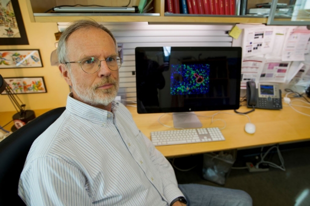
Roeland Nusse was awarded the 2017 Breakthrough Prize in Life Sciences for his contributions to the understanding a signaling molecule called Wnt.
Norbert von der Groeben
Nusse was awarded the $3 million prize for his contributions to the understanding of how a signaling molecule called Wnt affects normal development, cancer and the functions of adult stem cells in many tissues throughout the body.
“This is a complete surprise,” said Nusse, who is professor and chair of developmental biology. “My gratitude goes out to many people — my past and present postdoctoral scholars and graduate students and my former mentors have all contributed to the success of my research. The research and collaborative environment at Stanford and the long-term support from the Howard Hughes Medical Institute have also been fantastic. I see this award as a great honor for the entire community.”
The Breakthrough Prizes, initiated in 2013, honor paradigm-shifting research and discovery in the fields of life sciences, fundamental physics and mathematics. In total, about $25 million was awarded this year. A black-tie, red-carpet ceremony for the presentation of the prizes was held at the NASA Ames Research Center in Mountain View. The event was hosted by actor Morgan Freeman. The Grammy Award-winning pop star Alicia Keys provided entertainment.
“Roel’s pioneering work has provided deep insights into an essential molecular signaling pathway that controls normal embryonic development and adult tissue repair, and that contributes to cancer when it is not properly regulated. His work has served as a model for many others in our field and accelerated further studies of these critical processes,” said Stanford President Marc Tessier-Lavigne, PhD. “We are grateful that the Breakthrough Prize recognizes the work of scientific leaders who are inspiring others to pursue discovery that is truly transformative, benefiting all of humanity.”
His work has been the foundation of much of modern developmental biology.
Nusse’s interest in Wnt began in the 1980s as a postdoctoral scholar in the laboratory of Harold Varmus, MD, who was then on the faculty of UC-San Francisco. In 1982, Nusse discovered the Wnt1 gene, which was abnormally activated in a mouse model of breast cancer. He subsequently discovered that members of the Wnt family of proteins also play critical roles in embryonic development, cell differentiation and tissue regeneration.
“Roel has devoted his career to identifying one of the major signaling molecules in embryonic development, and clarifying its role in cancer development and in tissue regeneration,” said Lloyd Minor, MD, dean of the School of Medicine. “The importance of Wnt signaling in these processes cannot be overestimated. His work has been the foundation of much of modern developmental biology, and we are very proud of his contributions.”
Nusse’s more recent work has focused on understanding how Wnt family members control the function of adult stem cells in response to injury or disease. In 1996, he identified the cell-surface receptor to which Wnt proteins bind to control cells’ functions, and in 2002 he was the first to purify Wnt proteins — an essential step to understanding how they work at a molecular level.
“My work has shifted significantly over the years due to the influence of my Stanford colleagues, although it has always been focused on Wnt,” said Nusse. “When I arrived at Stanford, I was studying the involvement of the Wnt proteins in mouse development and cancer. I then switched to fruit flies, and then to the study of adult stem cells. Stanford has supported me during this evolution of my research career.”
Nusse’s lab is currently devoted to understanding how Wnt signaling affects the function of adult stem cells in the liver to help the organ heal after injury, as well as what role Wnt signaling might play in the development of liver cancer.
“The Breakthrough Prizes are a sign of the times,” said Nusse. “Together with the recently announced Chan Zuckerberg Initiative, they show how the wealth of Silicon Valley is now making an impact not just in the field of computer science, but also in biomedical fields. This is very exciting.”
The Breakthrough Prizes are a sign of the times.
Nusse is a member of the Ludwig Center for Cancer Stem Cell Research and Medicine at Stanford, of the Stanford Cancer Institute and of the Stanford Institute for Stem Cell Biology and Regenerative Medicine. He was awarded the Peter Debye Prize from the University of Maastricht in 2000. He is a member of the U.S. National Academy of Sciences, the European Molecular Biology Organization and the Royal Dutch Academy of Sciences. He is also a fellow of the American Academy of Arts and Sciences.
Seven $3 million Breakthrough Prizes — five in the life sciences, one in fundamental physics and one in mathematics — were awarded to 12 recipients. In addition, a special Breakthrough Prize in fundamental physics was awarded to the more than 1,000 researchers who proved the existence of gravitational waves in February of 2016.
By KRISTA CONGER
Krista Conger is a science writer for the medical school's Office of Communication & Public Affairs. Email her at kristac@stanford.edu.
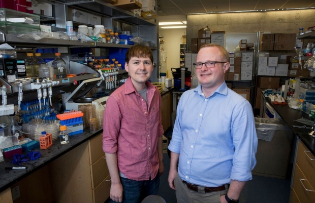
Prions can pass on beneficial traits, study finds
Researchers have found nearly 50 helpful prions in yeast and comparable proteins in humans, suggesting that this dreaded protein type can boost survival and plays a role in evolution.
Prion proteins, best known as the agents of deadly brain disorders like mad cow disease, can help yeast survive hard times and pass the advantageous traits down to their offspring, according to a new study by researchers at the Stanford University School of Medicine.

James Byers and Daniel Jarosz were part of a team that investigated how commonly traits in yeast are passed down by way of prion proteins instead of DNA.
Norbert von der Groeben
The study, published in the Oct. 6 issue of Cell and already available online, indicates that in yeast, and possibly other organisms, including humans, protein-based inheritance is more widespread than previously believed — and could play a role in evolution.
“In evolution there’s a paradox,” said Daniel Jarosz, PhD, assistant professor of chemical and systems biology and of developmental biology, who is lead author of the study. “We know that there are an extraordinary number of mechanisms that exist to protect the integrity of the genetic code and to assure that it’s faithfully passed on to future generations. But we also know that evolutionary success requires adaptability. How can you reconcile that need with the fact that the raw material for that innovation is really limited?”
Jarosz had a hunch that prions might be part of the answer. The new study suggests this could be the case.
How prions work
To understand his hunch, you need to know a bit about how prions work. Let’s consider this scenario in a bottle of beer on a hot summer day: When some of the yeast cells floating in your beer get stressed, in this case by the blazing sun, they begin producing large quantities of proteins called molecular chaperones — or proteins that help other proteins fold. These chaperones wrap around prions and fold them into a shape that kicks off a chain reaction of sorts. Other prions of its kind follow suit, using the original as a template.
“I sometimes liken it to a spreading fashion trend among teenagers,” said Jarosz. “Once it catches on with a couple of kids it spreads rapidly to other teenagers. But only teenagers.”
The upshot is that a single prion can quickly convert many others to assume the same shape — and since a protein’s shape dictates its behavior, that means the prion converts the other proteins’ behavior as well. Furthermore, when a cell divides, both new cells are likely to carry prion proteins that will continue to spur conversions. That means the offspring will also display the new behavior, a result of inheritance perpetuated not through the standard means of DNA but instead by way of proteins.
In the case of mad cow disease, a prion leads its normal brethren to fold in a way that leads to tissue damage in the brain and spinal cord of cattle. A human version of the disease, called variant Creutzfeldt-Jakob disease, can result from eating beef products from infected cattle.
They’re not all bad
But Jarosz knew that not all prions are bad. He had learned about a few beneficial ones from his time as a postdoctoral scholar at the Whitehead Institute for Biomedical Research in the laboratory of Susan Lindquist, PhD, a co-author of the study who has pioneered investigations of prions as a driver of inheritance. When Jarosz came to Stanford as an assistant professor in 2013, he began what would become a nearly three-year project to systematically assess an organism for prion-based inheritance. He chose yeast, he said, because researchers around the world have established the organism’s genetics and have developed comprehensive tools to analyze them.
“I wanted to know the breadth of protein-based inheritance across the yeast genome. Is it really so rare?”
With the help of several robots, his team overexpressed nearly every yeast gene, one by one, for 48 hours — triggering each gene to create 10 to 100 times more copies than usual of the protein designated by its code. Of the 5,300 genes they revved up, they found 46 made proteins that led to traits that remained heritable many generations after their expression had returned to normal. The traits were generally beneficial, such as resistance to temperature stress and anti-fungal drugs, or enhanced growth at high temperatures.
When they analyzed the proteins’ shapes, they discovered that few of them resembled what researchers had expected prions to look like. Most previously known prions fold in such a way that they pack tightly together and form long fibrils. The newly discovered prions lacked that trait, but many had others in common: They were strongly attracted to DNA molecules and they featured long, floppy “arms” able to fold in a wide variety of ways.
Non-Mendelian inheritance
Digging up unknown prions in yeast is less dangerous than you might think, said Jarosz. The efficiency of cross-species templating is very low for prion proteins. “This is probably one of the major reasons that mad cow disease wasn’t more widespread,” he said. “There is a safety concern with the human proteins. But luckily, simple procedures, like treating with bleach or soaking in sodium hydroxide, can render these protein conformations harmless.”
The team also found that the traits followed prionlike inheritance patterns. For example, unlike traits resulting from most genetic mutations, prion traits are dominant, and they don’t obey Mendel’s laws. Rather than being passed to half of all progeny in genetic crosses — as Mendel saw with his peas — prion-based traits are transmitted to every cell.
I sometimes liken it to a spreading fashion trend among teenagers.
Additionally, when the researchers temporarily inhibited chaperone proteins, the prion-based traits were permanently eliminated.
For the ultimate test, the researchers destroyed the DNA in the yeast cells carrying what they believed were prion-based traits, collected the remaining cell contents and introduced it into ordinary yeast cells. They found the traits were transmitted even though the cell’s DNA had been destroyed — indicating that proteins were transmitting the traits instead.
The researchers also found several human genes that would make proteins with similar characteristics. “These domains have been widely conserved across evolution, and several human homologs had the capacity to fuel protein-based inheritance,” they wrote in the study. “Our data thus establish a new and common type of protein-based molecular memory through which intrinsically disordered proteins can drive the emergence of new traits and adaptive opportunities.”
The work is an example of Stanford Medicine’s focus on precision health, the goal of which is to anticipate and prevent disease in the healthy and precisely diagnose and treat disease in the ill.
Other Stanford co-authors are James Byers and Richard She, both graduate students in Jarosz’s lab; David Garcia, PhD, postdoctoral scholar; Laura Lee, a graduate student formerly in Jarosz’s lab who is now working with professor of biology Dominique Bergmann, PhD; and former Amgen summer student Brayon Fremin, who is now a graduate student in the lab of Ami Bhatt, MD, PhD, assistant professor of medicine and genetics.
Researchers from the Whitehead Institute for Biomedical Research and MIT also co-authored the study.
The research was supported by the National Institutes of Health (grants R00GM098600, NIHDP2GM119140, T32GM007790, F32GM109680), the Ford Foundation, the Searle Scholars Program, the Sidney Kimmel Foundation, the David and Lucile Packard Foundation, the Howard Hughes Medical Institute, the Harold and Leila Mathers Charitable Foundation, the Eleanor Schwartz Charitable Foundation, the Broodbank Fellowship at the University of Cambridge, a Stanford Graduate Fellowship and the Stanford Summer Research Program/Amgen Scholars Program.
Stanford’s Department of Chemical and Systems Biology also supported the work.
By ROSANNE SPECTOR
Rosanne Spector is the editor of Stanford Medicine magazine for the medical school’s Office of Communication & Public Affairs. Email her at manishma@stanford.edu.